


In cell line development, a question that often surfaces only late in a project is: can this cell line be shown to originate from a single cell?
During the experiment, researchers may have completed cell isolation, culture, and screening, while also saving experimental images and operation records. When the project enters quality review, technology transfer, or submission preparation, the team often needs to revisit the early records and confirm several key points:
|
On Day 0, how can we confirm that there was truly only one cell at the target location? |
Determining monoclonality requires going back to the cell line establishment process itself. Demonstrating cell origin depends not only on the final outcome, but also on the records created as the experiment took place.
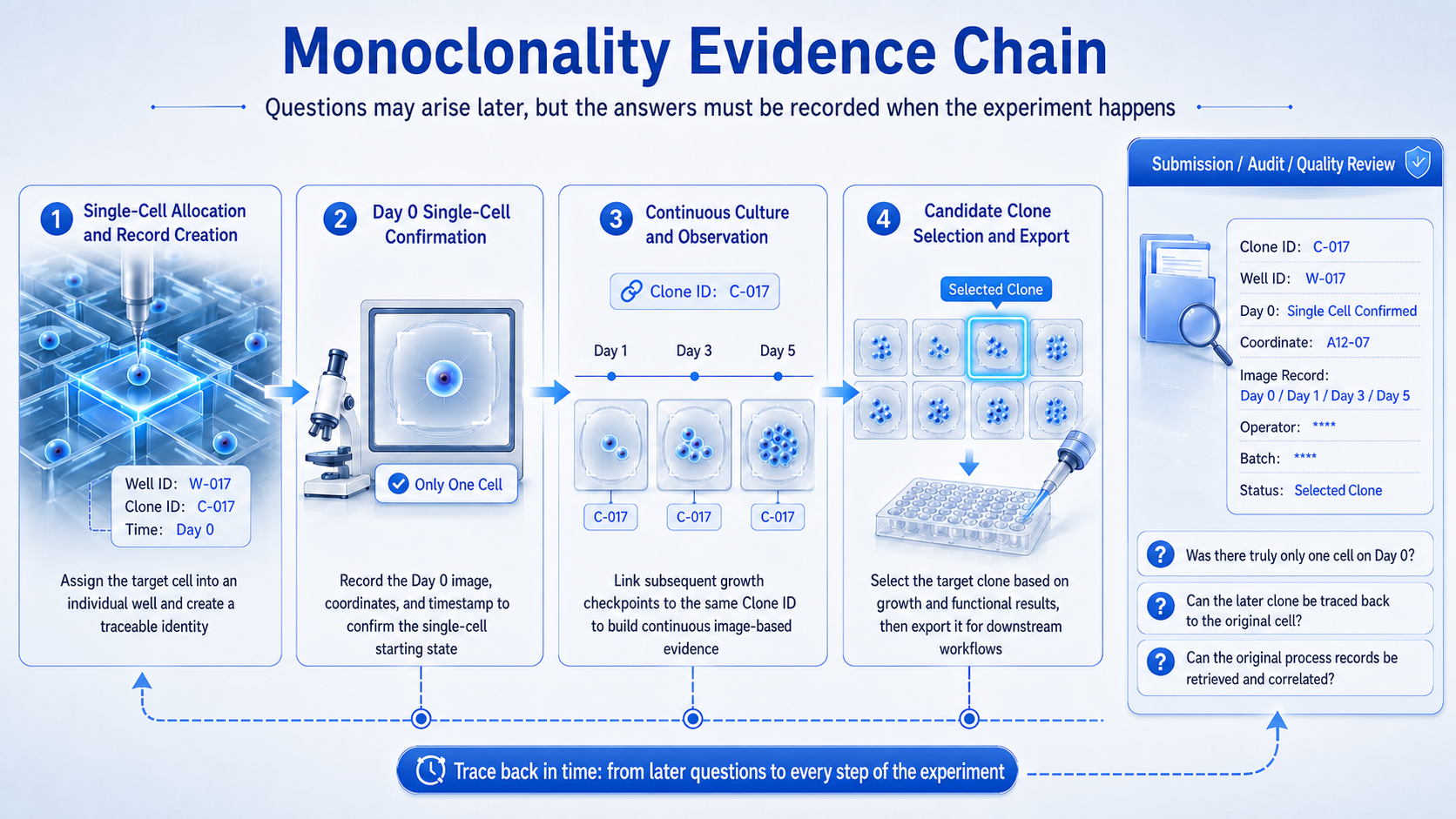
Evidence for monoclonality must be traced back to the cell line establishment process; later review depends on process records created during the experiment.
Monoclonality cannot be established by the final conclusion alone
The ICH Q5D guideline, Derivation and Characterization of Cell Substrates Used for Production of Biotechnological/Biological Products, issued by the International Council for Harmonisation of Technical Requirements for Pharmaceuticals for Human Use (ICH), addresses cell substrates used for production. It requires their origin, history, and generation process to be described, with relevant operation records retained throughout development. For production cell substrates established from cloned cell lines, the corresponding clonal origin and generation process must also be documented.
ICH Q5D does not prescribe a single technology pathway for demonstrating monoclonality. However, its requirements for documenting cell origin, generation history, and operation records make evidence retention an important part of cell line development.
Traditional limiting dilution methods typically use the inoculation concentration, dilution factor, and a statistical model to estimate the probability that a well originated from a single cell. This provides valuable statistical support, but it primarily answers one question: how likely is it that the well originated from a single cell?
During quality review, researchers also need to confirm whether the number of cells in the well was directly observed or supported by other evidence, and how the initial single cell can be linked to the clone that formed later.
Monoclonality evidence therefore usually comes from more than one result or method. Statistical inference, Day 0 images, cell position, sample IDs, operation time, subsequent culture records, and information associated with the experimental batch and operator can all form part of the evidence chain.
Statistical methods provide a probability basis, while process images and experimental records show more of what actually happened. They are not substitutes for one another; they should be used together according to the specific process, risk assessment, and quality system.
Why a Single Day 0 Image Is Not Enough
Day 0 imaging is an important part of monoclonality evidence. In this article, Day 0 specifically refers to the time point when a single cell is placed in the target well or culture chamber and an initial image is recorded. However, a single photograph does not automatically constitute complete proof.
With traditional limiting dilution, whether a complete and clearly visible single cell can be directly observed on the day of seeding depends on cell state, imaging conditions, and the observation method; it should not be treated as a default assumption. Regardless of the method used, the initial state must be assessed against defined imaging conditions, decision criteria, and review procedures to determine whether it truly supports a single-cell origin. For example, two cells may be adjacent, have overlapping boundaries, or fail to be fully identified because of differences in the focal plane. This assessment requires clear imaging conditions, decision criteria, and review procedures.
The initially observed single cell must then be linked clearly to the clone that forms later. Even if images are retained for both Day 0 and subsequent culture stages, it can be difficult to confirm that the records correspond to the same cell origin if the data from different stages do not share stable coordinates, well positions, sample IDs, or identity markers.
Finally, the records themselves must be trustworthy. When the image was created, which sample it represents, who performed the operation, whether the original record was retained, and whether it was processed afterward all affect whether the record can support future review.
The real value, therefore, is not that a photograph was taken. It is the record chain that starts with the initial single-cell state and remains continuously linked to the subsequent culture process.
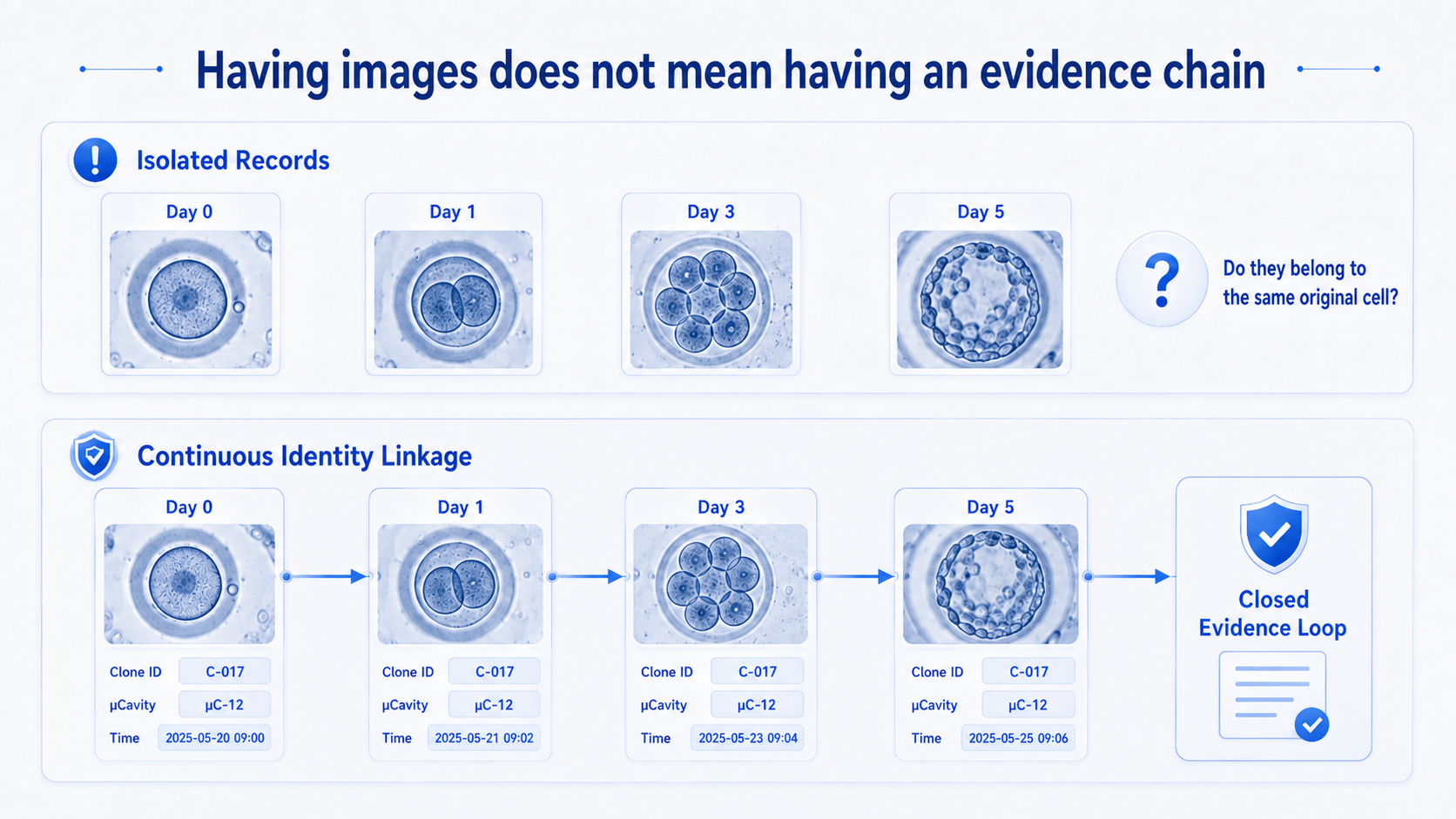
Comparison of an ineffective evidence chain and an effective evidence chain (conceptual illustration)
Enterprise-Grade Data Integrity Requirements
For cell line development records that must be retained and reviewed over the long term, data value depends not only on what the records contain, but also on whether the records can be trusted.
The ALCOA+ framework—Attributable, Legible, Contemporaneous, Original, Accurate, plus Complete, Consistent, Enduring, and Available—is commonly used to assess data integrity in regulated research and manufacturing environments. It asks whether data are attributable to a specific person, legible, recorded as the activity occurs, preserved in their original form, and accurate, while also remaining complete, consistent, enduring, and available throughout their lifecycle.
21 CFR Part 11, issued by the U.S. Food and Drug Administration (FDA), provides technical controls and governance requirements for the use of electronic records and electronic signatures in relevant settings. Part 11 is not a "certification label" that can be attached to an instrument. It focuses on whether an organization has established appropriate technical measures, management processes, and validation activities around the use of electronic records and electronic signatures. Whether a system can support a specific compliance context therefore usually depends on two layers: the technical capabilities provided by the system itself, such as user identity management, access control, and record protection; and the validation, procedures, and quality system established by the user for the intended use. This two-layer structure means that equipment suppliers and customers carry different but connected responsibilities.
In practice, organizations typically need to answer a series of specific questions:
Which data qualify as electronic records requiring controlled management?
How does the system ensure that experimental time, equipment information, and operator information are recorded together?
What access and operation permissions does each user have?
Can every modification be traced to a specific person, time, and reason for the change?
Does the system provide a complete audit trail?
How are images, sample information, and operation records linked?
How are data stored, backed up, and retrieved?
When a review takes place in the future, can the experimental context and operation process be reconstructed?
These questions cannot be solved by relying on a single instrument, nor can they be solved by a single standard operating procedure. They require coordinated support from software controls, instrument use, personnel management, validation activities, and the quality system. An instrument can provide part of the technical foundation, but it cannot replace the user's overall responsibility for data integrity and the quality system.
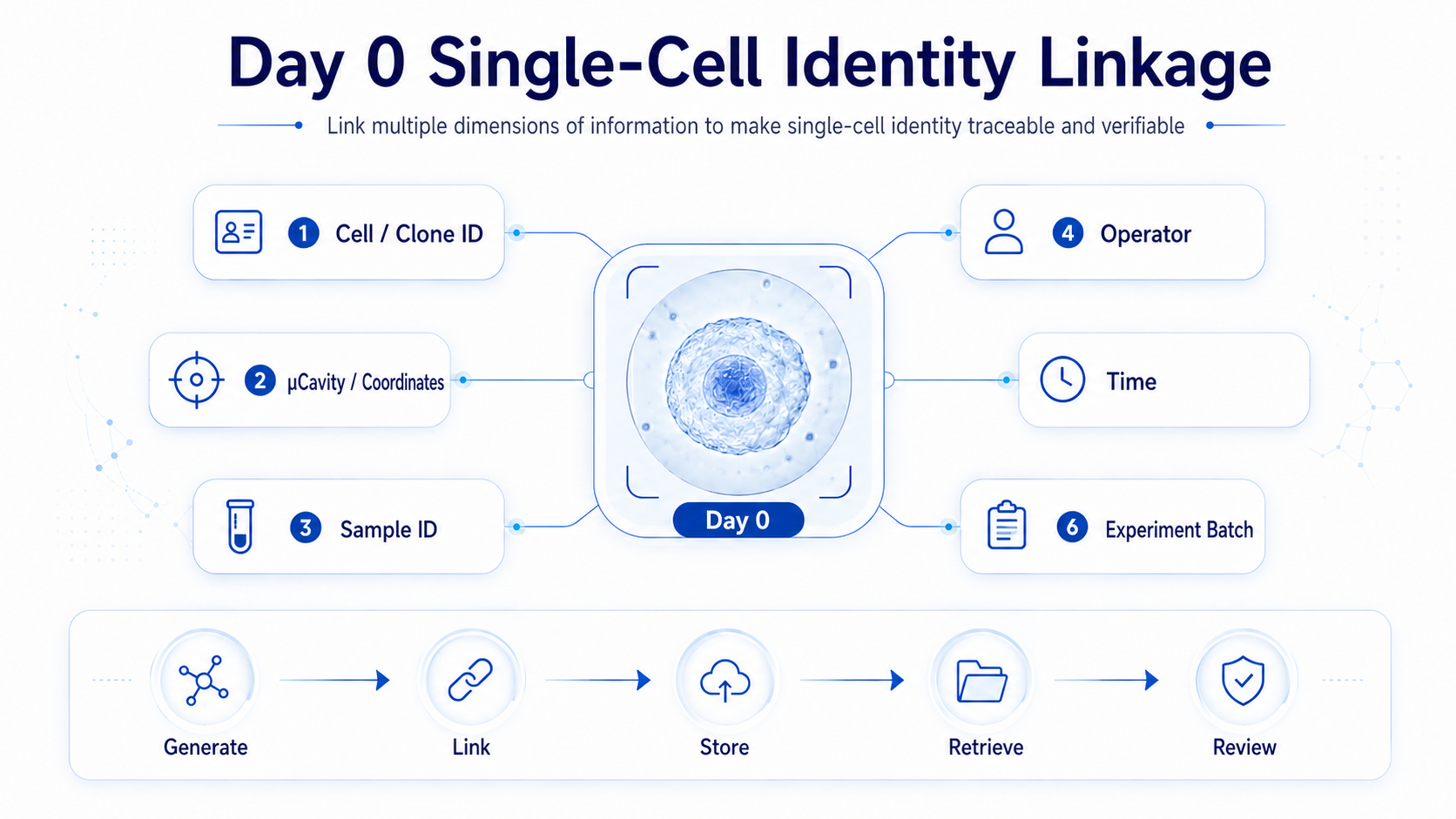
Linking Images with Identity, Time, Sample, and Operational Context
How OptoBot® 1000 Contributes to the Single-Cell Evidence Chain
OptoBot® 1000 is designed for cell line development and single-cell screening. It combines optoelectronic tweezers (OET), a microfluidic chip, and imaging modules to identify, select, manipulate, and observe cells during culture under microscopic observation.
OET projects programmable light patterns onto a photoconductive layer to form light-induced virtual electrodes. These electrodes create a non-uniform electric field inside the chip, enabling non-contact cell manipulation through dielectrophoretic forces. While observing cell state, researchers can select and move a target cell, allowing both which cell was selected and how the operation occurred to be observed and recorded together.
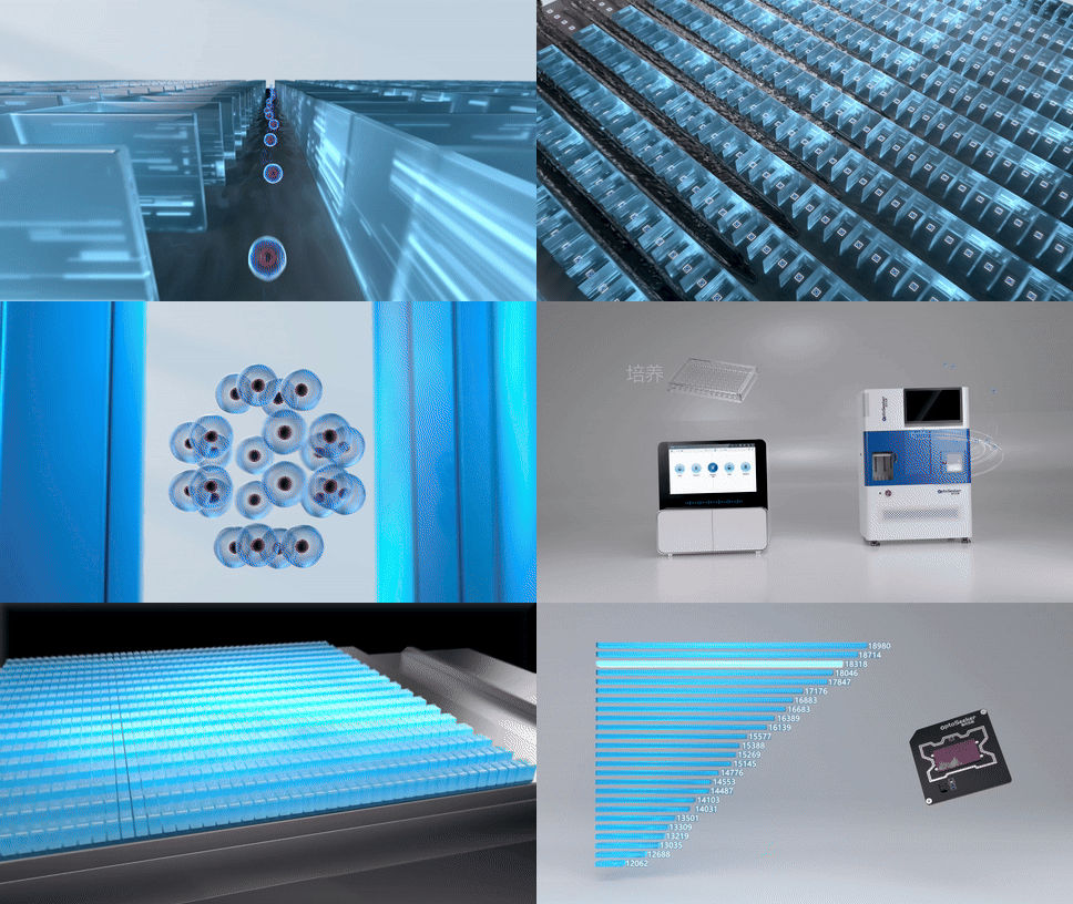
OptoBot® 1000 enables precise cell loading, culture and expansion, secretion analysis, export, and single-cell record creation with identity numbering.
In the cell line development workflow, Day 0 is not only a confirmation of the single-cell state; it is also the starting point for subsequent identity tracking.
OptoBot® 1000 can link the Day 0 single-cell image to subsequent clone-growth images through the same coordinates or ID. During a later review, researchers can follow this identity relationship to view the initial single-cell state at the target position and the growth process that followed.
Compared with images that are independent of one another at different time points, this continuous linkage can reduce a common problem: every stage may have a record, yet there is no proof that the records belong to the same cell origin.
In addition, OptoBot® 1000 software provides data-management and security functions aligned with relevant FDA 21 CFR Part 11 requirements. It supports tiered user permissions, identity binding, electronic signatures, and audit trails, linking relevant operations to specific user identities and providing a technical basis for experimental-record attribution and permission management.
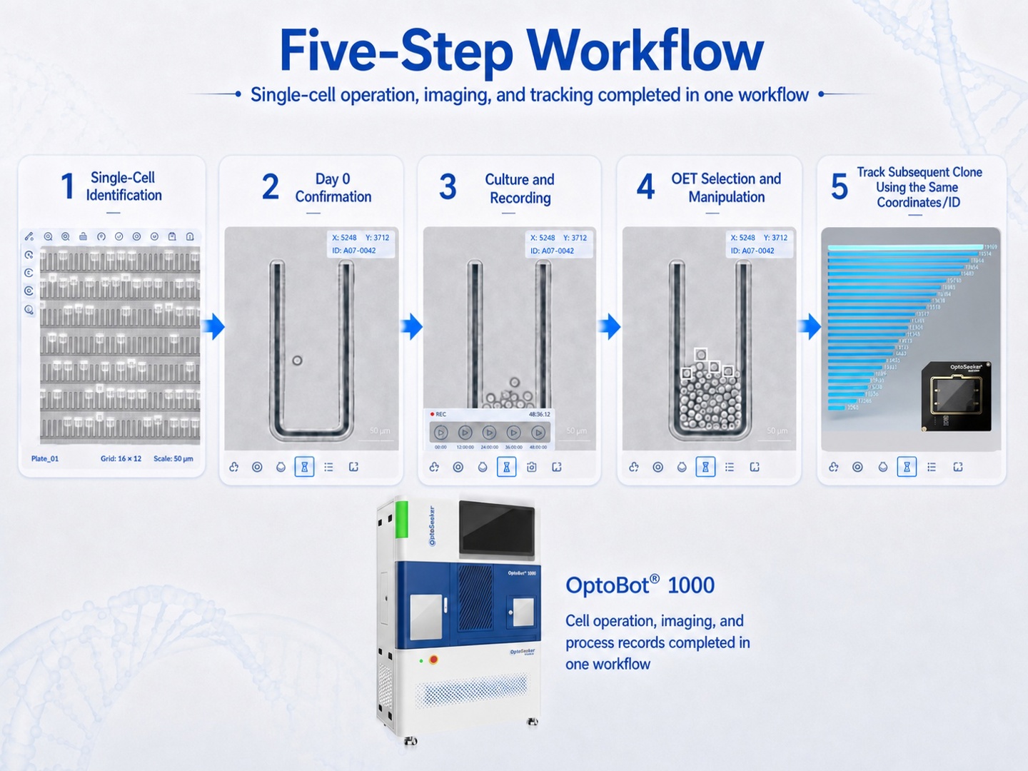
Conceptual illustration of the OptoBot® 1000 process-evidence workflow — cell operation, imaging, and process records are completed within one workflow
|
As a high-throughput single-cell optofluidic sorter that integrates optoelectronic tweezers, a fully automated microfluidic chip, and OptoMind® AI software, OptoBot® 1000 automates the workflow from cell loading, single-cell isolation, and on-chip culture to multidimensional characterization, functional screening, and live-cell export. It also provides real-time imaging, data archiving, and traceability analysis throughout the experiment. In antibody discovery, it can validate the function of antibodies secreted by individual B cells and link the results to sequencing data. In cell line development, it can screen monoclonal cells with high viability and high productivity. In T-cell function analysis, it can link functional phenotypes with molecular information across multiple dimensions, including cytokine secretion, surface markers, and dynamic killing. |
Let Every Clone Return to Its Starting Point
Ultimately, demonstrating monoclonality is not only about answering whether a well or microcavity may have started from a single cell. It is about whether, when researchers revisit a cell line, they can follow the existing records back to its initial state.
Through visual single-cell identification and manipulation, Day 0 image recording, subsequent clone-growth observation, and continuous identity linkage, OptoBot® 1000 enables cell selection, operation, and culture to generate corresponding records within one workflow.
Together, these records form more than an isolated single-cell photograph. They form a traceable history built around a specific clone.
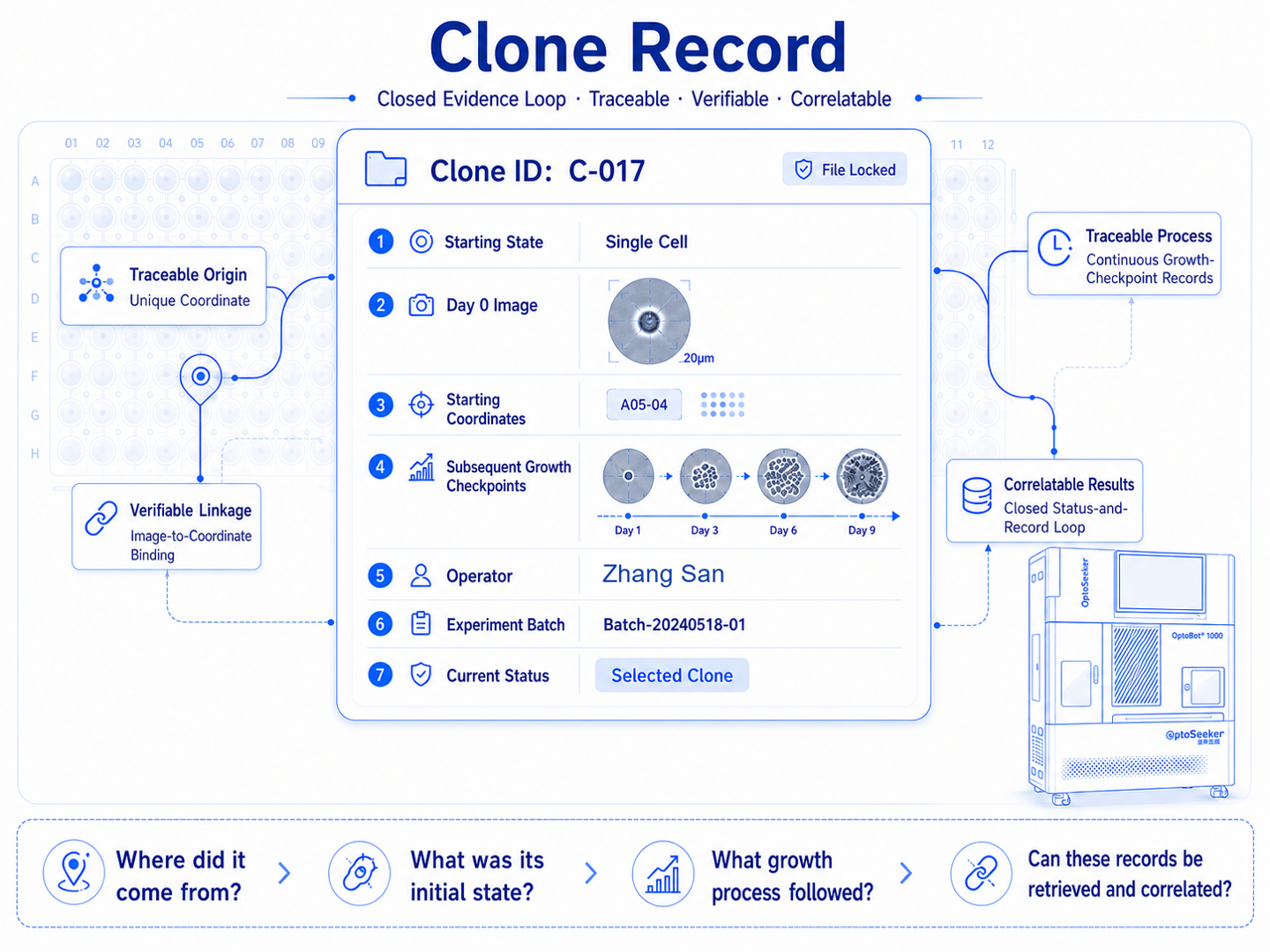
Single-Cell Record Card (conceptual illustration)
OptoBot® 1000 does not replace the user's responsibility to build the complete quality system, nor can a single instrument alone produce a final monoclonality conclusion. What it provides is a clearer process-evidence foundation: the initial single cell, the clone that forms later, and the related records generated during the experiment no longer remain isolated data points.
Only when these data remain continuously linked around the same starting cell can a monoclonality assessment become an evidence chain that can be reviewed, explained, and retained over the long term.