

Today's AI is already remarkably capable.
It can read papers, summarize findings, propose experimental hypotheses, and even offer judgments close to those of researchers in many specialized tasks. Yet in life science, the most important question is often not on the screen but under the microscope: in a complex cell sample, where is the one cell that is truly worth selecting and tracking further?
Because many answers in life science are not found in text.
They are hidden in the real experimental setting. In antibody discovery, for example, the challenge is not only to identify an individual B cell that secretes a high-potential antibody, but also to match the fluorescence response to the exact cell and recover, export, and analyze the B-cell receptor (BCR) sequence of the candidate cell.
This is where the OptoBot®1000 Series High-Throughput Single-Cell Optofluidic Sorting System from OptoSeeker Biotech creates value. Batch acquisition of B-cell sequences and transcriptomic information cannot, by itself, determine whether the antibodies secreted by a given cell have the target function. OptoBot®1000 uses the limited in vitro activity window available during B-cell culture to read functional phenotypes—such as antibody secretion and antigen binding—at the single-cell level first, and then selectively export hit cells for sequencing.
1. Biological Answers Are Found in Real Cells
AI models can readily explain how antibody-secreting cells (ASCs, such as plasmablasts and plasma cells) or activated B cells produce antibodies. But reasoning alone cannot determine, in a real sample, which cell secretes an antibody with the most desirable binding or functional characteristics and is worth advancing into development.
That answer is not contained in any single paper. The functional phenotype of a particular cell in a specific sample cannot be inferred from literature or a model alone; it still needs to be measured and validated experimentally.
A sample may contain thousands or even more B cells. They may look similar, yet their functional behavior can differ dramatically. The candidate cells truly worth advancing may be only a very small fraction of the population.
If researchers can see only a population-average signal, rare but high-value cells can easily disappear in the background. Finding them requires working at single-cell resolution: seeing and distinguishing cells one by one, then tracking them through the downstream workflow.
2. The Hardest Step: Seeing the Function—and Retrieving the Blueprint
In an antibody discovery experiment, the most exciting moment is often not the end of the experiment, but the moment when a signal first appears.
In the microscope field, fluorescence around certain B cells begins to intensify. In the data table, several candidate readouts are clearly above background. From the results, the experiment seems to have captured a valuable lead.
But when the lead moves into downstream development, the questions begin:
|
Which cell produced this signal? |
In a traditional workflow, functional testing, cell sorting, culture, sequencing, and bioinformatic analysis are often distributed across different platforms. One experiment may produce a fluorescence signal, one sorting run may produce a cell population, and one sequencing run may produce sequence files. But if these data cannot be traced reliably back to the same individual cell, the single-cell evidence chain becomes fragile.
In other words, screening cannot stop at "seeing fluorescence." A screening process with real industrial value must recover the specific cell that generated the signal, establish where it was transferred, and create a traceable upstream record for downstream sequencing and confirmation.
3. Operating Cells with Light: From Experimental Technique to Standardized Workflow
So how can a cell only a few micrometers in size—and highly fragile at the same time—be moved with precision?
One of the underlying technologies used by OptoSeeker Biotech is optoelectronic tweezers (OET).
Under an applied AC electric field, projected light patterns change local conductivity in the chip's photoconductive layer, creating movable virtual electrodes and a non-uniform electric field. Cells are positioned, moved, or captured through dielectrophoretic forces. Here, light modulates the electric field; unlike traditional optical tweezers, it does not directly hold cells through optical gradient forces.
Combined with a microfluidic system, this allows researchers to complete key steps—including cell positioning, identification, movement, on-chip testing, and export—on a single chip.
The value of OptoBot®1000 is therefore not only that it makes "operating cells with light" possible. It also turns a task that once depended heavily on individual experience into a standardized experimental workflow for real-world research and development.
Here is how the system completes this workflow in three steps:
Step 1: Give Every Candidate Cell a Traceable Identity
After the sample enters the OptoTrap Chip®, OptoBot®1000 can identify target cells under the microscope and distinguish single cells, doublets, debris, and abnormal particles.
The system then uses OET-based optofluidic manipulation to guide qualified cells into individual microcavities.
This step may look basic, but it determines whether all subsequent data can be trusted. The system can establish a tracking identity for a specific microcavity and its candidate cell, continuously linking its position, images, and experimental state.
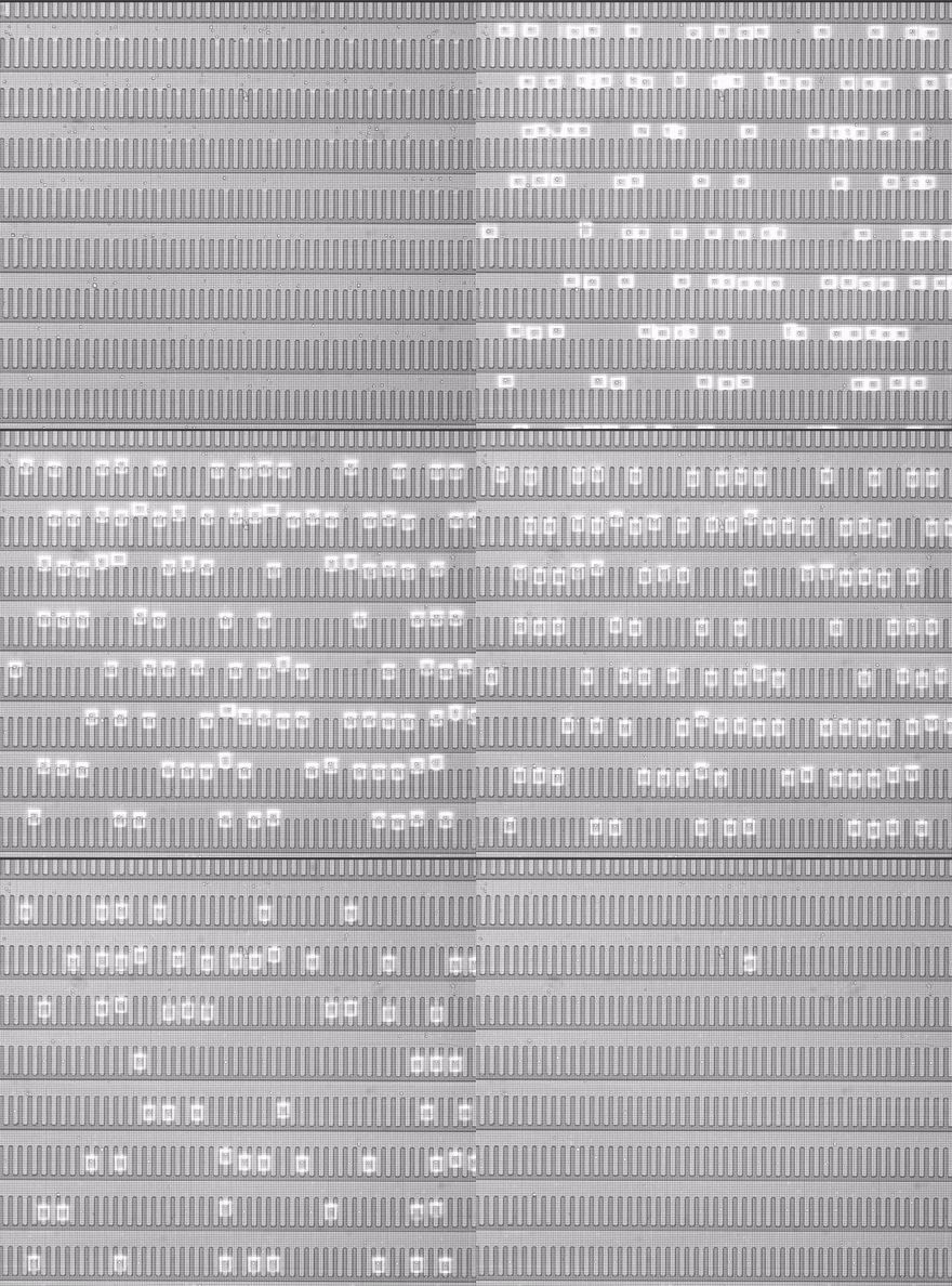
Figure 1. OET-based optofluidic manipulation guides a single cell into the target microcavity, establishing a spatial foundation for subsequent on-chip testing and tracking.
For antibody discovery, high throughput means that more candidate B cells can enter the same single-cell tracking framework, more cells can be compared, and more functional readouts can be collected in parallel. When sample quality, assay performance, and screening criteria are comparable, expanding single-cell coverage can increase the opportunity to discover rare candidates.
Step 2: Read Functional Performance Directly on the Chip
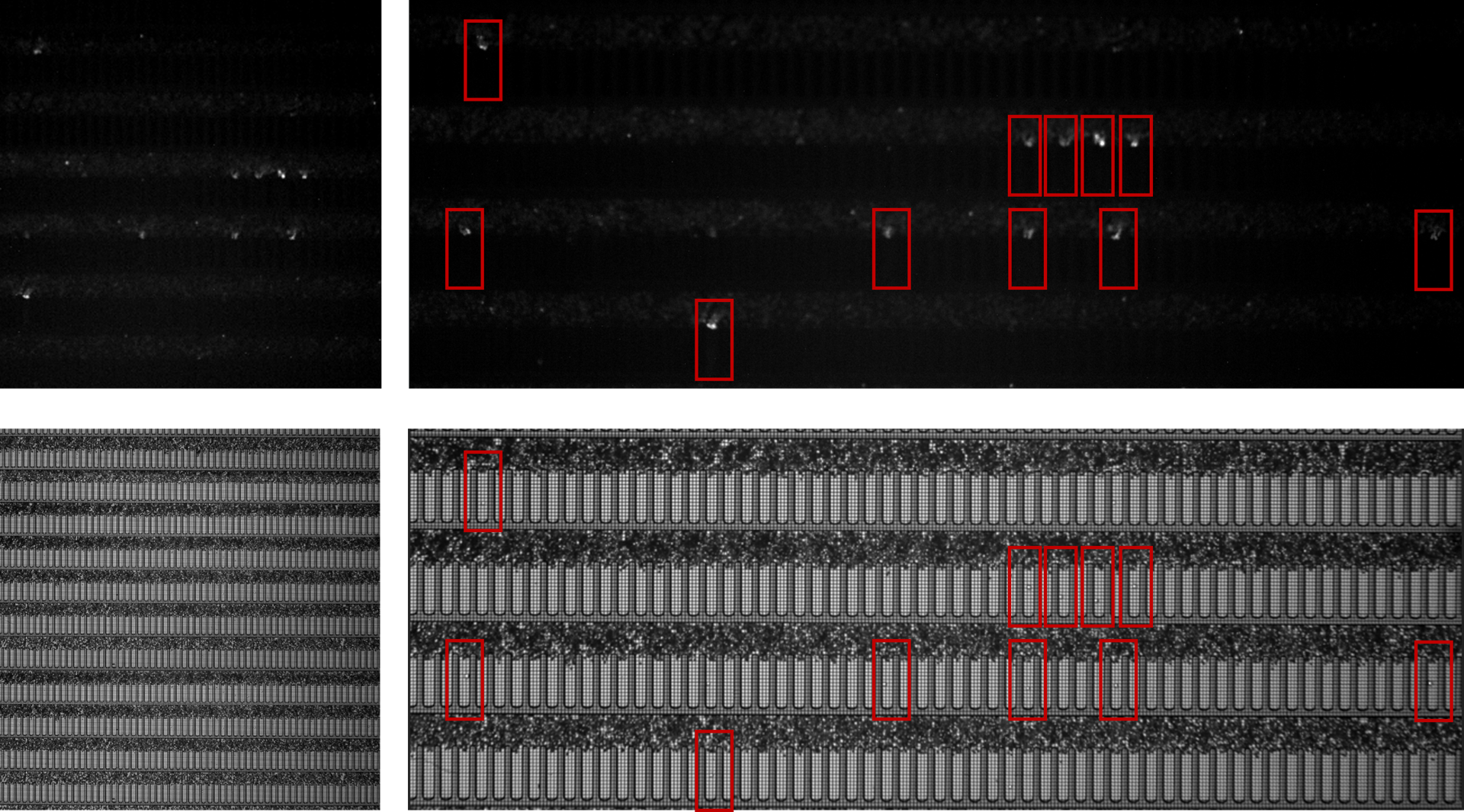
Figure 2. The system can identify candidate cells in brightfield and fluorescence views and link them to microcavity position, image records, and functional readouts.
The fluorescence changes, secretion signals, and morphological states read by the system are no longer just signals in a field of view. They become functional measurements linked to a specific cell, a specific microcavity, and a specific time point.
With an appropriate fluorescence assay, OptoBot®1000 can read information such as antibody secretion, binding to a target antigen, and cell state. Further assessment of specificity, blocking or neutralizing activity, or affinity requires corresponding counter-screening, functional assays, or quantitative binding experiments.
As the assay progresses, fluorescence around some microcavities gradually increases, while other microcavities remain at low or background levels. It is important to note that fluorescence is an experimental readout generated by a specific assay system. Its intensity and rate of change are influenced by multiple factors and cannot, on their own, be equated with antibody affinity or functional activity. The system can combine original microscopic images, fluorescence intensity, time-series changes, and cell morphology to score and rank candidate cells.
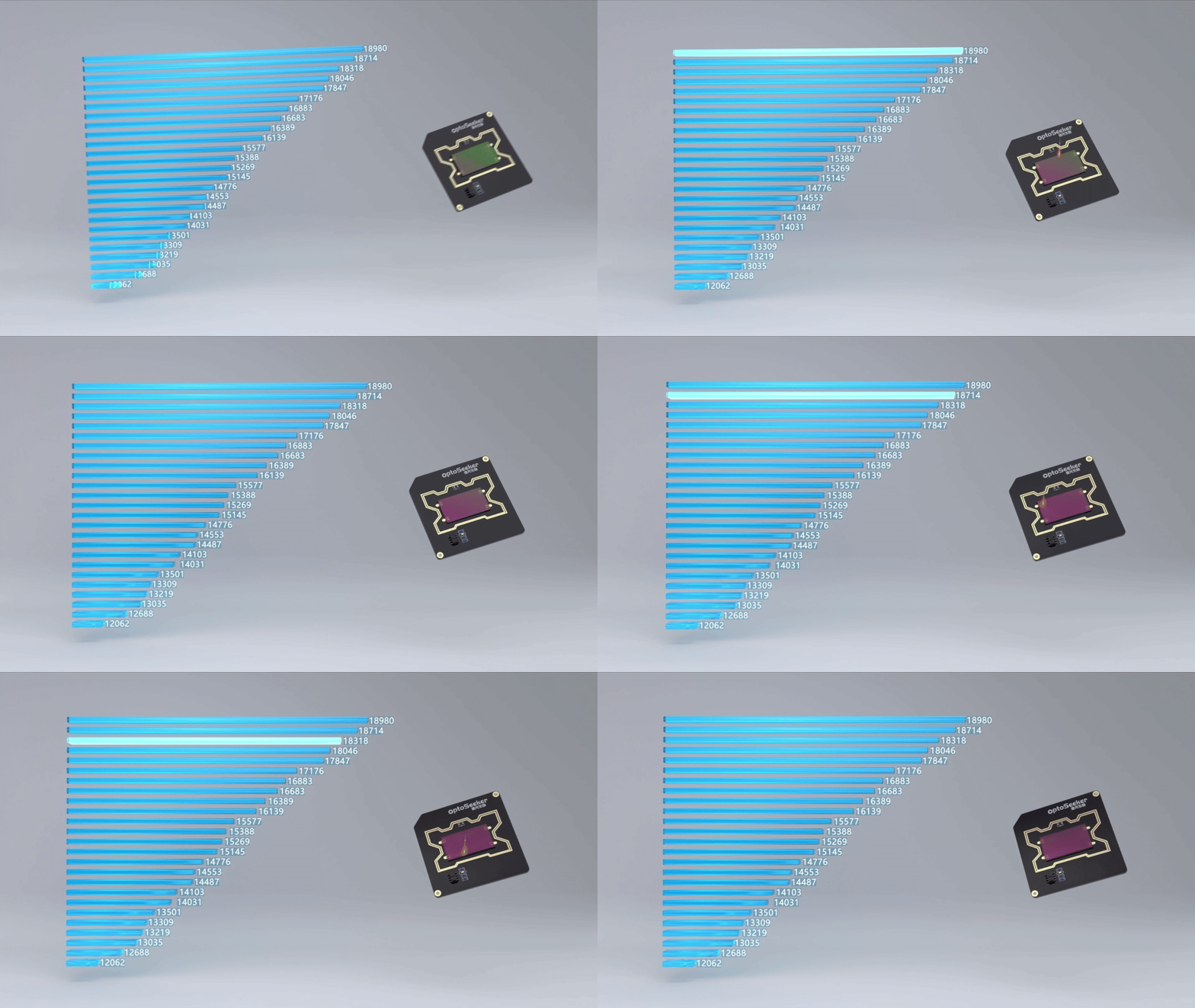
Figure 3. Candidate cells can be ranked by functional readouts and entered into an export workflow for downstream sequencing or culture.
This "function-first" screening strategy allows researchers to observe how cells actually perform in a functional context before deciding which cells are worth exporting, sequencing, and confirming. For antibody discovery, this is closer to the information needed for development decisions than simply relying on a population-average signal.
Step 3: Bring the Hit Cell Back into the Experimental Workflow
A functional signal is only the entry point.
A bright fluorescence spot or an attractive time-course curve has real value for further development only when the corresponding cell can be relocated and exported.
One of the key capabilities of OptoBot®1000 is to relocate a candidate cell from its original position on the chip and link the export path, destination well, and operation records to the cell's unique identity.
Researchers can then carry out amplification, library preparation, sequencing of immunoglobulin transcripts, acquisition of paired heavy- and light-chain variable-region sequences—that is, B-cell receptor (BCR) sequences—and expression confirmation while maintaining a clearer link to the individual-cell origin.
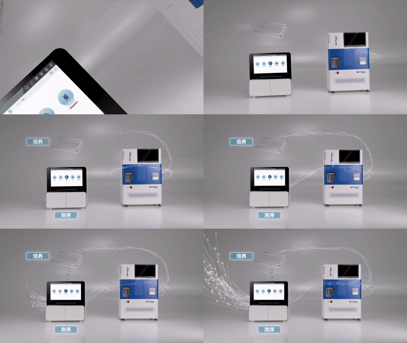
Figure 4. Integrating single-cell operation, on-chip testing, and data recording provides the foundation for bringing candidate cells back into the downstream experimental workflow.
Complete single-cell genotype–phenotype pairing is still affected by factors such as downstream amplification efficiency, library-preparation bias, bioinformatic quality control, and contamination exclusion. These are challenges shared across the industry. The value of OptoBot®1000 is that, when single-cell origin, export well position, and downstream sample barcodes are kept consistent, and after amplification, pairing, and contamination-control QC, on-chip phenotypic readouts can be linked to the heavy- and light-chain sequences of the candidate antibody.
4. Beyond Antibody Discovery: Expanding the Boundaries of Single-Cell Exploration
The core capabilities of OptoBot®1000—including precise single-cell positioning, on-chip culture, dynamic functional testing, and downstream-oriented cell export—are not limited to antibody discovery.
In cell line development (CLD), the system can record single-cell seeding and clone formation, providing traceable imaging evidence for single-cell origin. It can also be used to measure and sort high-producing cell lines on the chip.
In research related to cell and gene therapy (CGT), the system can be used to observe dynamic interactions between T cells and target cells, while recording cell-state and function-related signals according to the assay design.
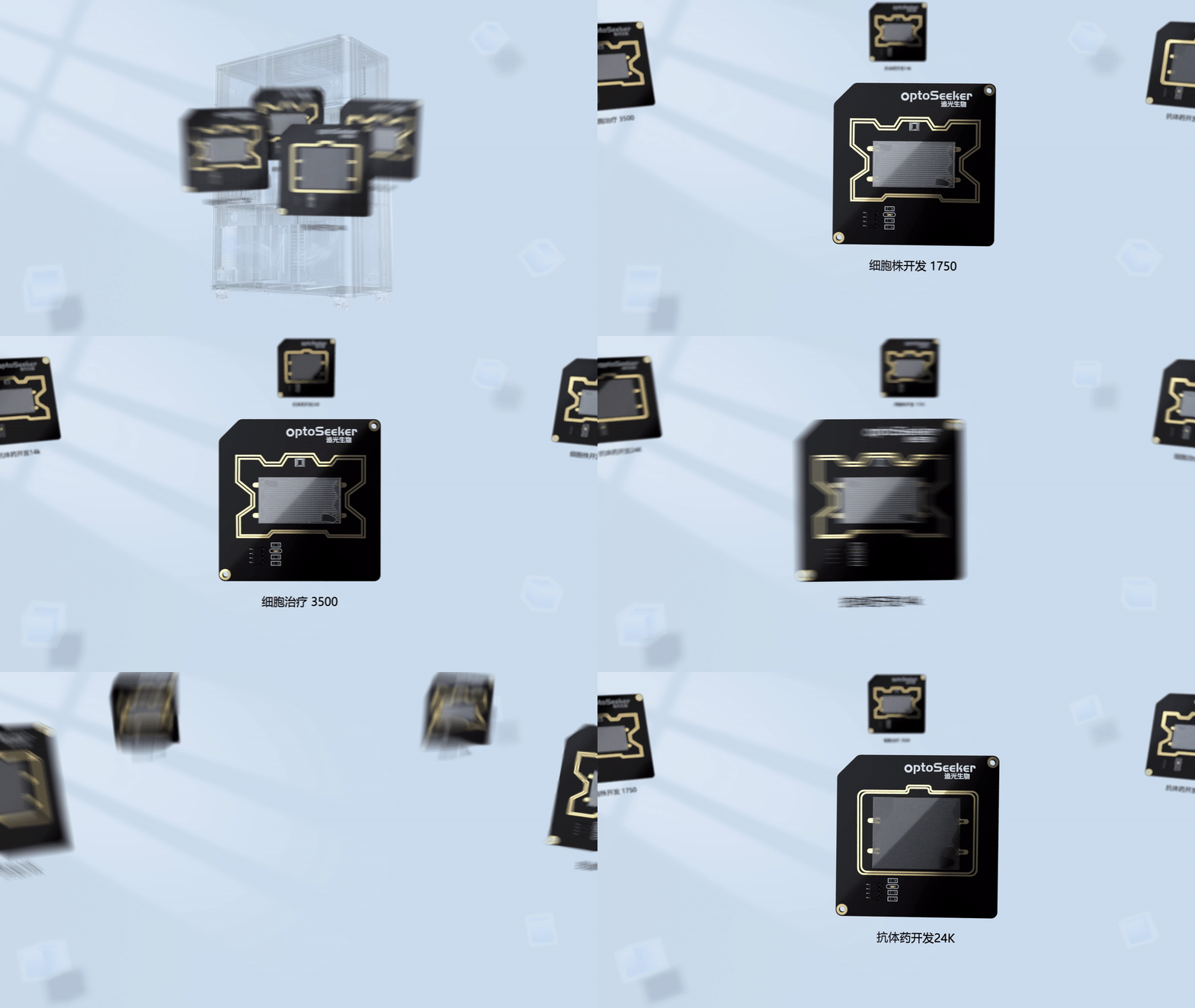
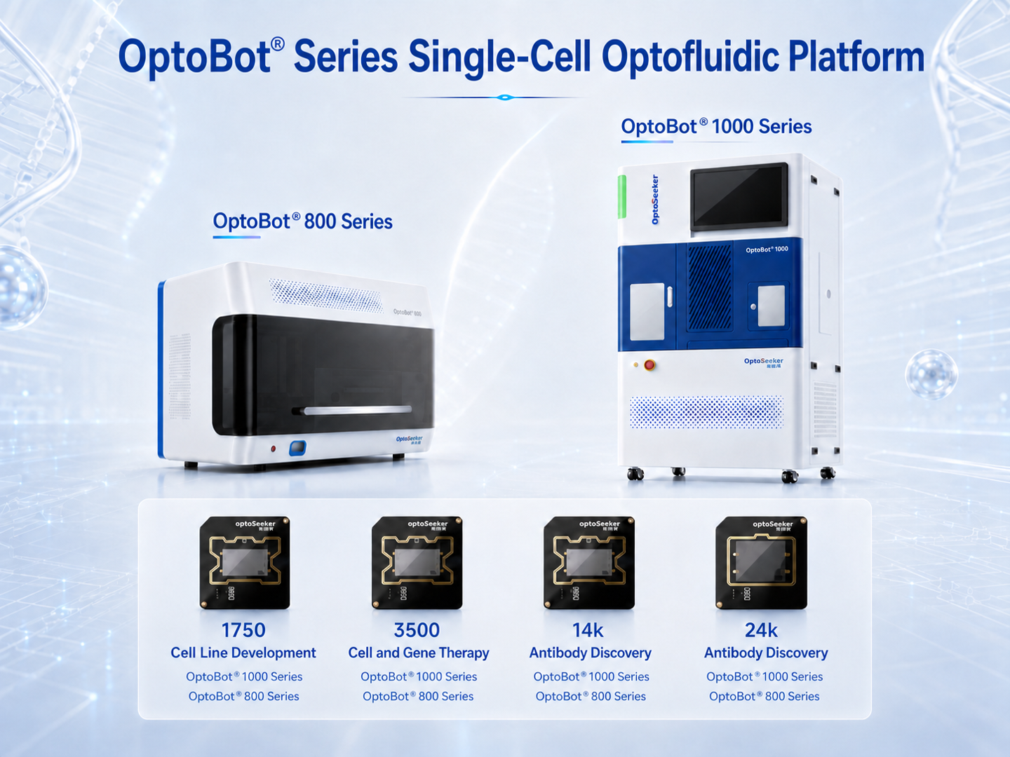
Figure 5. OptoBot®1000 can be paired with OptoTrap Chip® devices in different specifications to support single-cell applications including antibody discovery and cell line development.
Ultimately, all of these applications ask the system for the same core capability: precisely confirming and extracting the one truly valuable cell from a complex, heterogeneous population.
5. From Instrument to AI for Science Infrastructure
When a system connects single-cell positioning, functional testing, candidate ranking, targeted export, and downstream analysis, what it records is no longer limited to "what genes this cell has."
More importantly, researchers can trace what experimental operations the cell underwent, what functional responses it produced, why it was selected, and which downstream analysis and validation path it ultimately entered.
OptoMind® software already provides AI-assisted cell identification and data-analysis capabilities. Looking further ahead, as single-cell experiments continue to accumulate structured images, operation records, phenotypes, sequences, and downstream validation data, AI may gain access to a data foundation that is closer to the real experimental process. It could then move beyond analyzing existing results to participating in experimental understanding and research-and-development decisions.
The significance of OptoBot®1000 goes beyond completing a single cell-sorting run. OptoSeeker Biotech aims to build it, through coordination among software, hardware, chips, and experimental workflows, into an experimental platform connecting the real world of cells with computational analysis. One end is oriented toward living cells, performing positioning, observation, manipulation, and export. The other is oriented toward the data system, accumulating single-cell evidence that can be tracked, re-validated, and analyzed further.
Conclusion: Make Every Cell Worth Tracking Traceable
AI has taught machines to read and think. But the last mile of life science still depends on observation, manipulation, and validation in real experiments.
The industrial value of a high-throughput single-cell instrument cannot be judged only by how vivid its fluorescence images are or how large its nominal throughput numbers look. The real test is whether it actually closes the data links most likely to break in the discovery workflow.
From single-cell positioning and in situ functional testing to multidimensional candidate ranking and physical export of hit cells, OptoBot®1000 connects these critical steps.
When a cell has a traceable identity from the moment it enters the chip, each flash of functional activity is no longer an experimental data point with no clear origin. What researchers deliver is no longer an ambiguous "candidate mixture," but the specific location, functional record, and physical path to downstream experiments for each cell.
Leaving clear evidence for every cell worth tracking and preserving a traceable origin for downstream development: this is the key answer OptoBot® 1000 provides at the scale of single-cell manipulation.