


In life-science research, finding a “positive” cell is often only the starting point for the next round of work.
In antibody discovery, identifying candidates that bind the antigen is not enough. The next questions are: How strong is the affinity? Do the candidates deliver the blocking or other functions required by the program?
In cell line development, teams face large numbers of candidate clones and need to determine how they grow and secrete, and which ones are worth expanding further.
In T-cell functional research, surface markers or population-averaged data alone cannot answer every question: Which cells effectively kill target cells? Do cells that secrete more cytokines also show stronger killing activity? Can these behaviors be linked to molecular information from the very same cell?
Across these research settings, richer single-cell functional information is needed to support decisions and move the cells of interest into the next stage of research.
OptoSeeker OptoBot®1000 connects single-cell manipulation, culture, functional assays, screening, and live-cell recovery, helping researchers see how cells actually perform earlier in the workflow.
| Function-Guided Cell Selection: OptoBot®1000 Capabilities and Applications |
From a Single Cell to an Informed Selection
OptoBot®1000 is a high-throughput single-cell optofluidic platform built around optoelectronic tweezers (OET), integrating microfluidic chips, multimodal imaging, automated control, and the OptoMind® software system. It brings precise single-cell manipulation, in situ culture, longitudinal observation, functional assays, and live-cell recovery onto one platform, delivering an integrated workflow that conventional instruments and standalone methods cannot readily match.
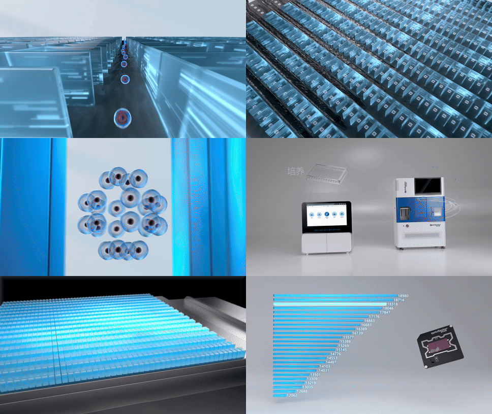
OptoBot®1000 supports precise cell loading, culture and proliferation, secretion assays, targeted export, and per-cell record creation with unique identity assignment.
Gentle, contact-free manipulation with optoelectronic tweezers is a key advantage of the platform.
The system uses programmable light patterns to reshape the local electric field in the chip and guide target cells, enabling contact-free, gentle capture, isolation, and transfer of individual cells. This approach reduces the risk of damage from mechanical contact and repeated pipetting, helping preserve cell viability for subsequent culture, functional testing, and expansion.
After sample pretreatment and workflow setup, cells are loaded into the chip and precisely guided into microchambers by OET. Microchannels continuously support medium replacement and reagent exchange, while the microchambers provide a relatively stable space for culture and observation, allowing the same cell to be followed over time for changes in growth, secretion, and function.
OET-based optofluidic manipulation gently guides individual cells into target microchambers, establishing the spatial basis for subsequent on-chip assays and tracking.
Around the same live cell, the system can perform multiple rounds of functional assays on-chip while continuously observing changes in cell state.
Bright-field and multi-fluorescence-channel imaging can be combined with assay steps configured for the experimental objective, linking cell morphology and growth state with functional signals to provide multidimensional evidence for selection.
Multiple chip formats support high-throughput screening across different applications. The throughput comes from the microchamber architecture: a single microchamber is approximately 100,000 times smaller in volume than a well in a standard 96-well plate, while one chip can contain thousands to tens of thousands of such chambers - up to 24K parallel chambers for antibody discovery. Cells remain physically separated, and secreted signals rapidly accumulate in the confined volume, improving assay sensitivity.
The overall process can therefore be summarized as:
Single-cell loading → On-chip culture and observation → Secretion and functional assays → Target selection → Live-cell recovery
Once cells of interest are identified, the system can export them directionally into downstream containers such as well plates for further expansion, sequencing, or other analyses. Images and data from loading, culture, assays, and export can all be recorded and reviewed, preserving the link between each cell’s functional performance and downstream research.
The result is a target live cell with functional information, a traceable process record, and the ability to continue into downstream research.
Why OET: Gentle Manipulation, Continuous Observation, On-Demand Recovery
The right single-cell screening tool depends on the type of information the study needs to obtain.
Fluorescence-activated cell sorting (FACS) excels at rapidly analyzing large numbers of cells and sorting them based on fluorescence and related signals. Droplet microfluidics uses large numbers of isolated droplets to support parallel assays and high-throughput screening.
OptoBot®1000 instead emphasizes linking functional assays, continuous observation, and targeted recovery around the same live cell. Researchers can make selections based on multiple dimensions of cell behavior during culture and testing, then move the selected cells into downstream studies.
For projects that compare antibody function, prioritize cell clones, or study dynamic T-cell behavior, this means gaining decision-relevant evidence earlier and focusing downstream work on the candidates that matter most.
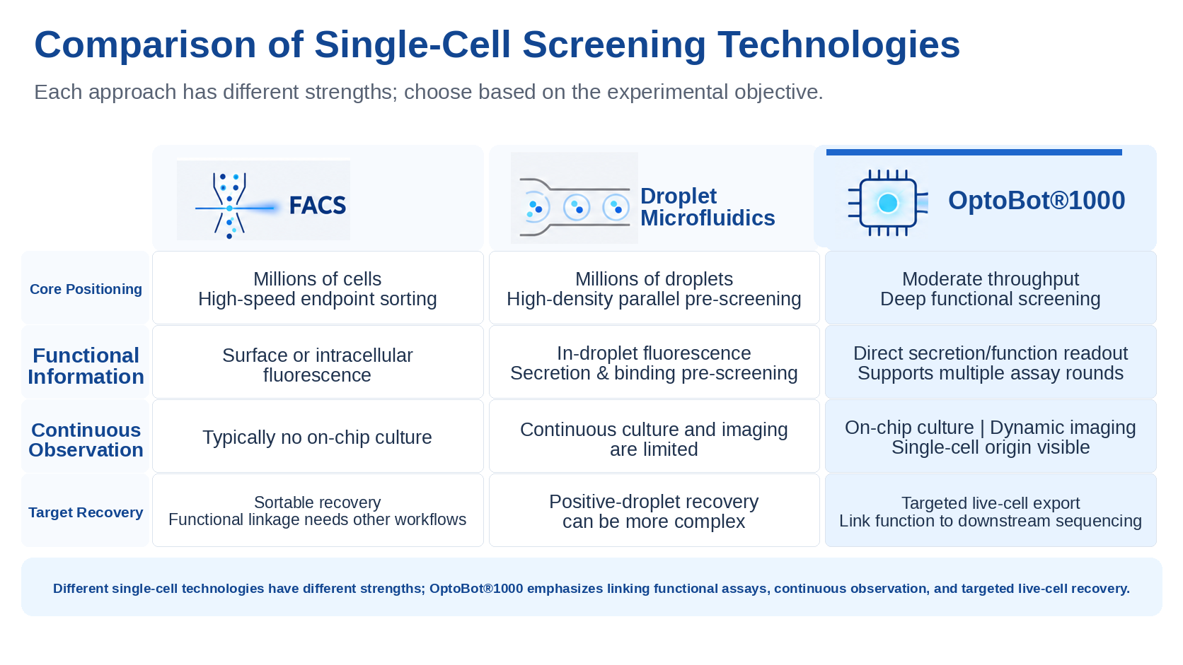
Comparison of Three Single-Cell Screening Approaches
Application 1 | Antibody Discovery: Bring Functional Evidence into Candidate Selection Earlier
Antibody discovery requires finding, among many candidates, antibodies that match the development objective. Beyond antigen-binding signals, affinity, cross-reactivity, blocking activity, and other functional properties can determine whether a candidate is worth advancing.
OptoBot®1000 moves these assays upstream to the single-cell stage.
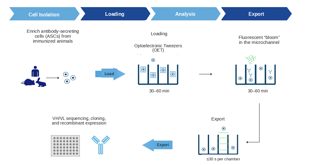
OptoBot®1000 Antibody Discovery Workflow
Once an individual B cell enters a microchamber, its secreted antibodies can be analyzed using detection reagents delivered through the microfluidic channel. In an antigen-specificity assay, for example, interactions between the secreted antibodies and the detection system generate an imageable fluorescence signal. A gradually forming fluorescent “bloom” near the microchamber opening provides a readout for identifying the corresponding antibody-secreting cell.
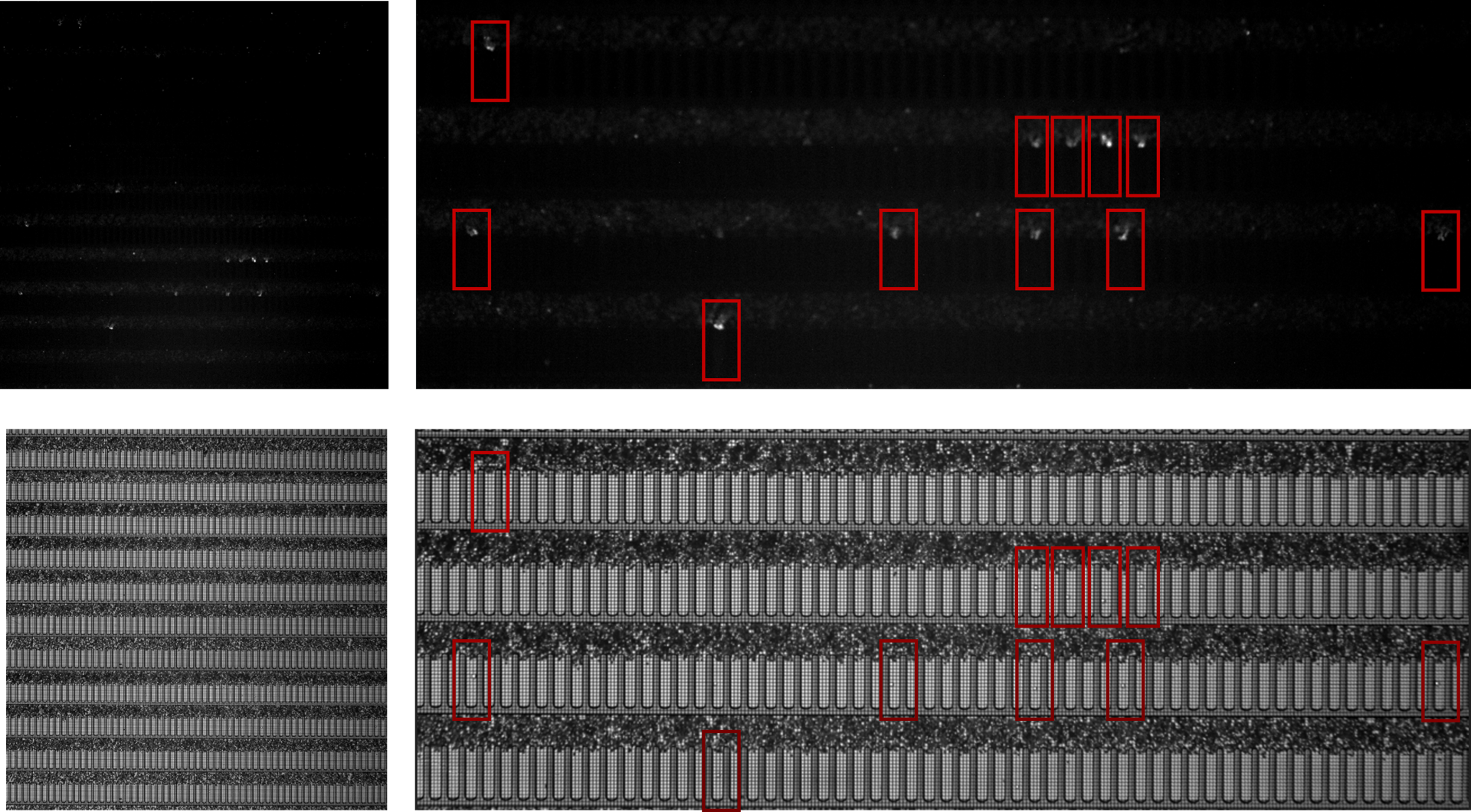
After B-cell-secreted antibodies interact with the assay reagents, imageable fluorescence enrichment can form near the microchamber opening, enabling identification of target antibody-secreting cells.
The platform can perform multiple assays over multiple rounds according to project requirements:
1. Antigen-specificity assay: identifies cells that secrete antibodies binding the target antigen.
2. Relative affinity scoring: combines antigen-binding and antibody-secretion-related signals to compare candidate binding performance under the specified assay conditions.
3. Project-relevant functional assays: use assays such as cross-reactivity, ligand blocking, and internalization to further narrow the candidate pool.
As assay information accumulates, researchers can select cells based on the target function and export them for downstream antibody sequencing, recombinant expression, and deeper validation.
Candidates entering the downstream workflow therefore already carry preliminary functional evidence relevant to the program objective.
In compatible nanoliter-scale assay formats, secretion signals can be observed locally at an earlier stage, with the full set of assays completed within one day.Combined with multidimensional screening, this helps reduce the downstream expression, testing, and analysis resources spent on poorly matched candidates.
Application 2 | Cell Line Development: Identify Clones Worth Expanding Earlier
Cell line development requires identifying, from many candidates, the clones most suitable for continued culture and validation. OptoBot®1000 brings single-cell origin, early growth, secretion assessment, and targeted export into one workflow, providing a stronger evidence base for early-stage prioritization.
First, establish where each clone came from.
From the moment a single cell is loaded, the platform continuously records its growth and proliferation and maps those records to the export destination, providing image and process evidence for monoclonal origin.
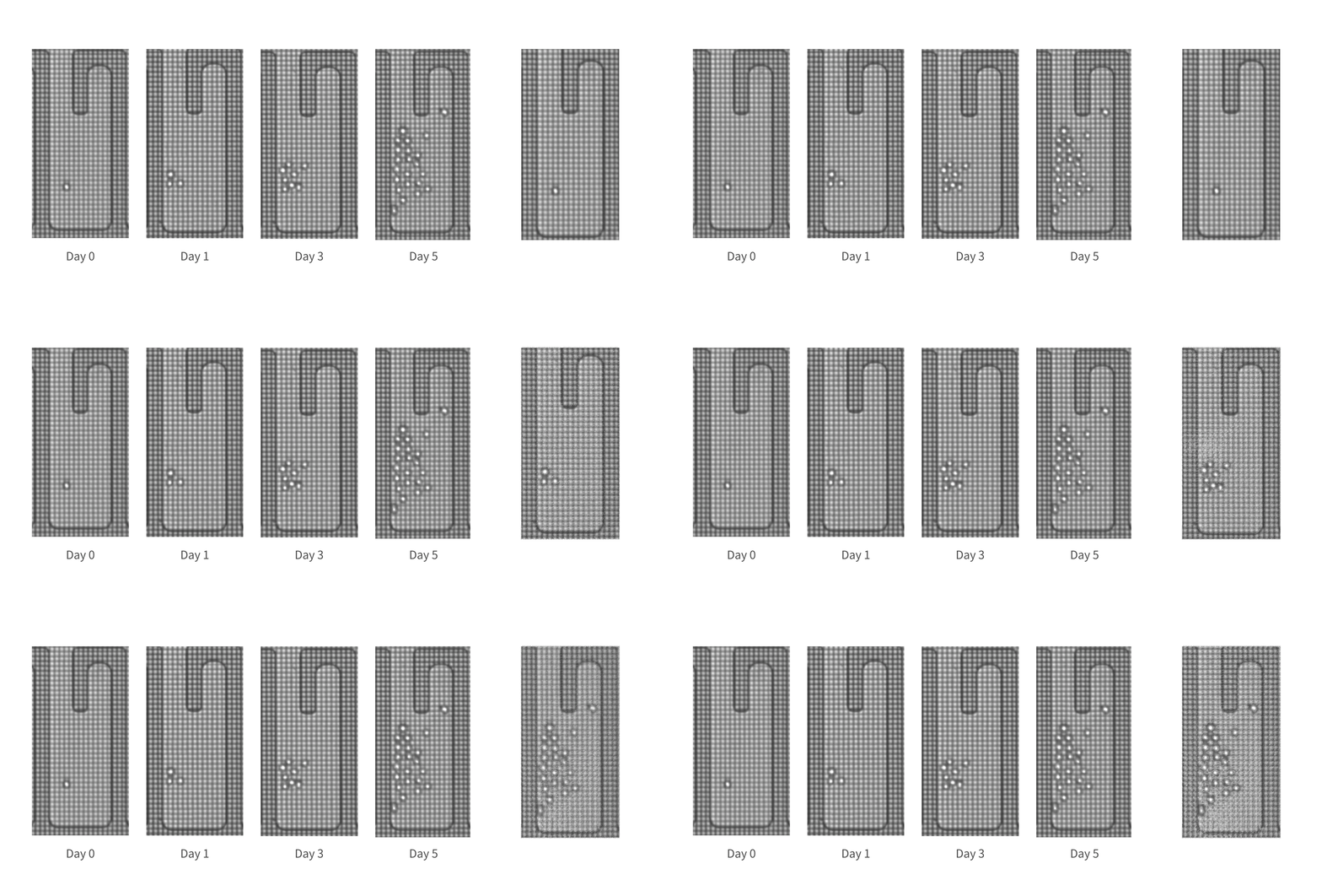
Recorded proliferation of cells within microchambers
Next, select clones by combining growth and secretion performance.
Higher total output may be associated with faster cell proliferation. By jointly analyzing cell counts, growth dynamics, and secretion signals, researchers can compare candidate clones more comprehensively and assess their production potential.
The platform can also incorporate aggregation and other quality-related assays as required by the project, providing additional screening information before clones are expanded further.
Selected target clones are then exported directionally into well plates for continued expansion, stability testing, and process development.
The more evidence-based the front-end screening, the more downstream culture, testing, and scale-up can focus on the most promising candidates.
Automated workflows also reduce some repetitive operations and sample-transfer steps, helping limit operator-to-operator variation and maintain more continuous experimental records.
Application 3 | T-Cell Functional Analysis: Connect Cell Identity with Actual Performance
Within a T-cell population, individual cells can display very different functional behavior. Understanding that heterogeneity requires linking multiple dimensions of observation back to the same cell.
OptoBot®1000 supports combined analysis of cytokine secretion, target-cell killing, and surface-marker expression to characterize individual T cells from multiple angles.
After T cells and target cells are placed together in microchambers, continuous imaging can capture their interactions and dynamic killing behavior. Corresponding assay systems can then be used to analyze cytokine secretion and surface markers.
These data can help researchers investigate questions such as:
1. Do T cells that secrete more cytokines also show stronger killing activity?
2. How are different surface-marker profiles related to secretion and killing behavior?
3. Under the relevant experimental design, can a given T cell serially kill multiple target cells?
Once cells of interest are identified, the platform can export them into well plates for TCR sequencing or single-cell transcriptomic analysis, enabling further investigation of the relationship between functional phenotype and molecular characteristics.
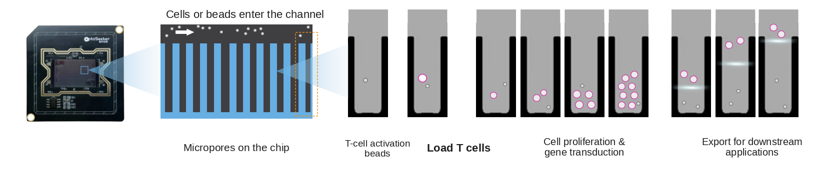
The platform can evaluate individual T-cell function across multiple dimensions and output quantitative functional
kinetic parameters and time-series data, directly linking cellular “functional phenotypes” with genotype/transcriptome profiles.
From “How does this cell behave?” to “Which molecular features are associated with that behavior?”, functional assays can be connected directly to downstream molecular analysis.
From Chips to Software: Adapting the Platform to Your Research Needs
Different studies place different demands on cell-culture space, screening scale, and assay format. With multiple chip configurations and editable experimental workflows, OptoBot®1000 helps researchers organize experiments around specific objectives.
Chip selection starts with the research need.
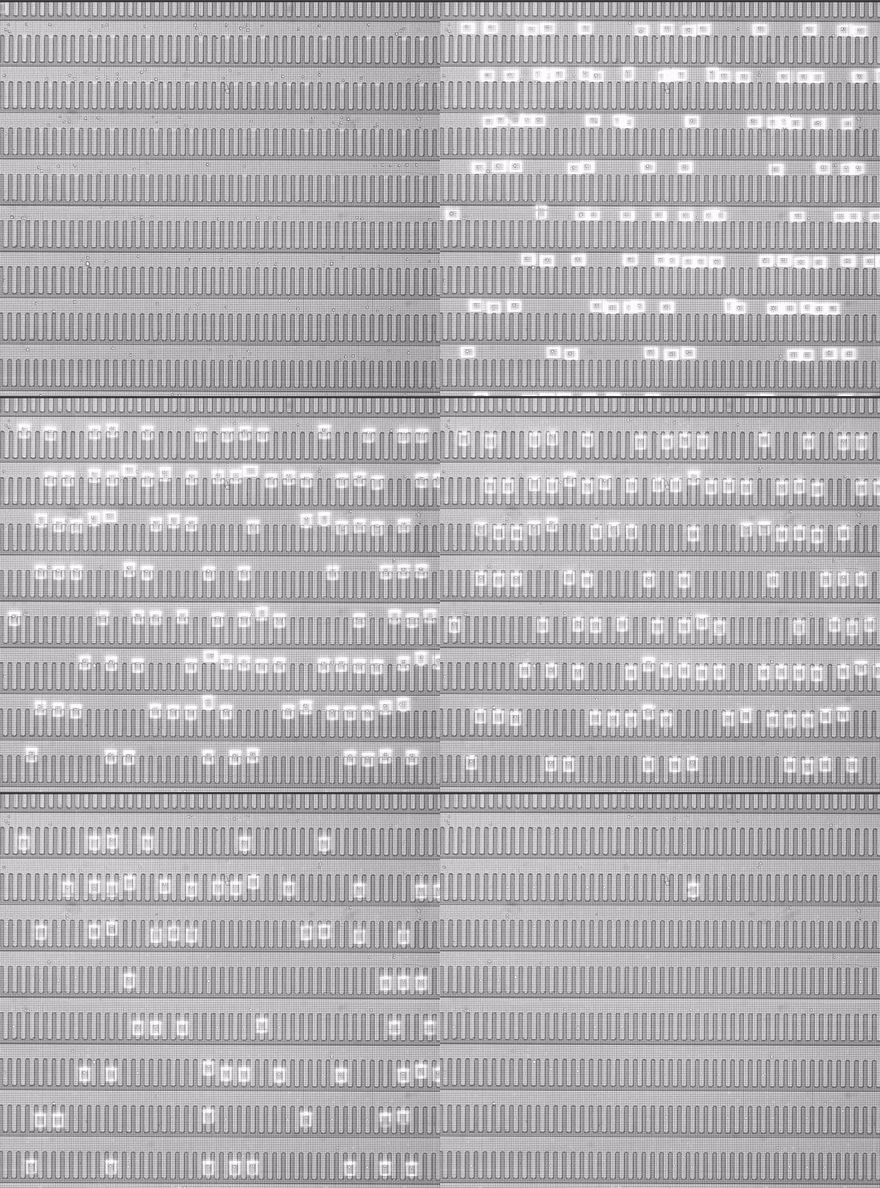
In cell line development, researchers focus on sustained culture of monoclonal cells, early growth, and secretion performance. The 1,750-chamber chip, with approximately 1.70 nL per chamber, provides relatively ample space for single-cell culture and subsequent clone export.
In cell and gene therapy applications, the 3,500-chamber chip, with approximately 0.74 nL per chamber, balances the needs of single-cell manipulation, culture, and functional analysis.
For antibody discovery, 14K and 24K higher-throughput chips support larger-scale single-cell isolation, on-chip culture, and functional screening, helping researchers identify cells with the desired function from large candidate pools.
|
|
|
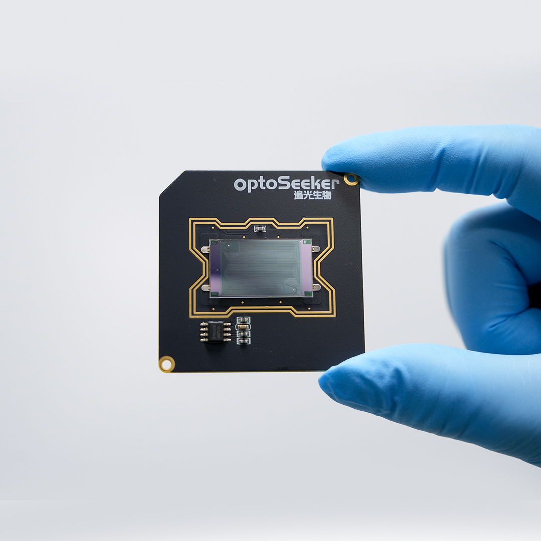
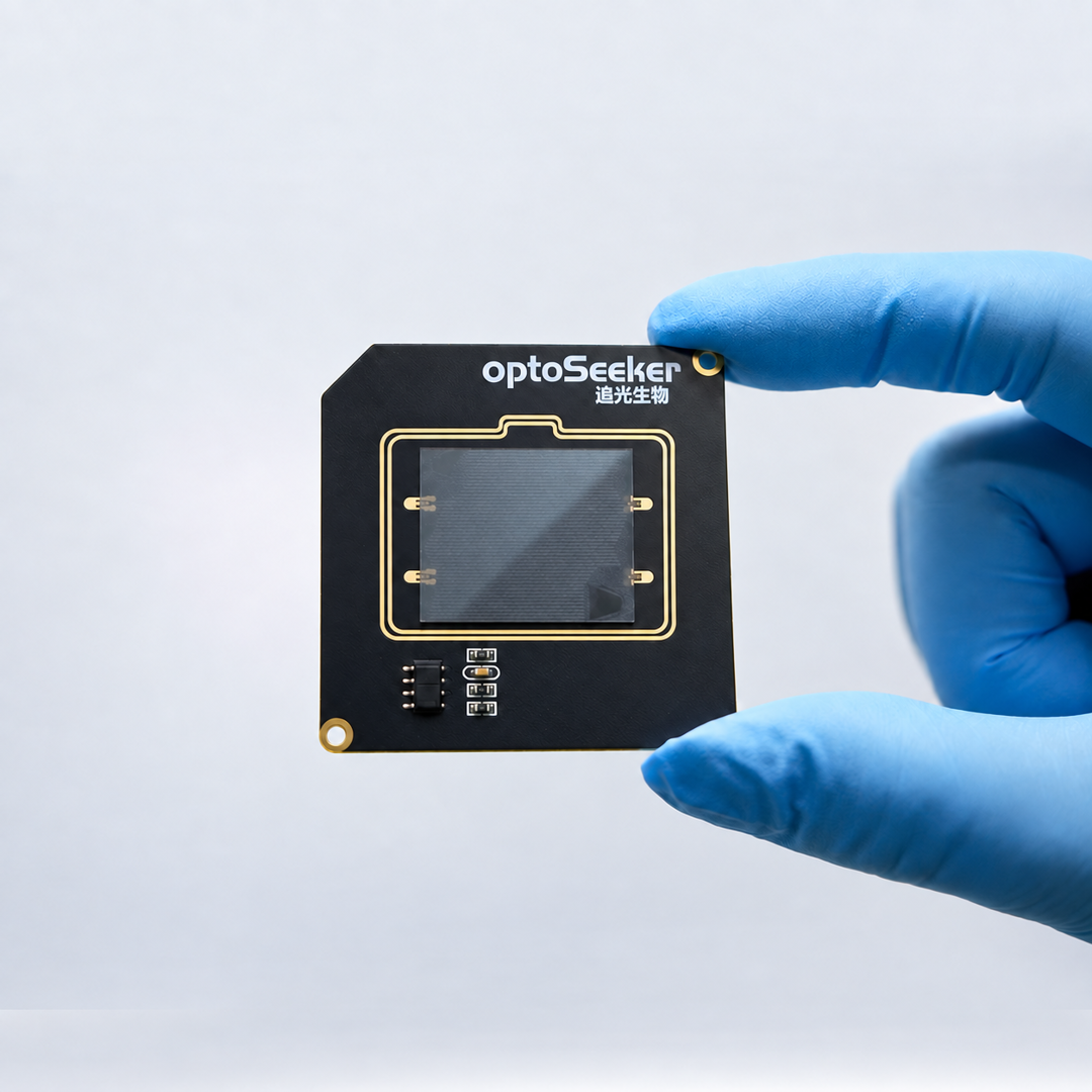
OptoBot®1000 uses microfluidic chips with different specifications for cell line development, cell and gene therapy, and antibody discovery (left: 1,750/3,500/14K chips; right: 24K chip).
Choosing the right chip requires balancing culture space, screening scale, and downstream assay requirements. On this basis, OptoBot®1000 uses OptoMind®software to organize individual experimental steps into a coherent workflow and convert on-chip cell information into actionable screening criteria.
OptoMind®provides built-in workflow templates and supports flexible editing based on experimental requirements. During execution, AI assists with cell recognition and path planning, while image-analysis tools automatically quantify cell number, position, morphology, and predefined fluorescence signals, reducing repetitive manual interpretation.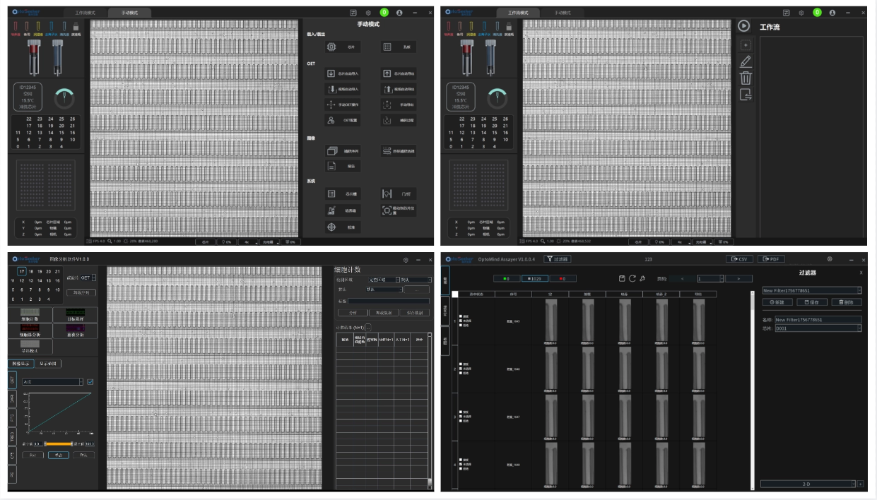
OptoMind® connects workflow execution, image analysis, and data review, making target-cell culture and assay processes filterable, reviewable, and traceable.
OptoMind® also provides a comprehensive data-management and security framework that meets relevant FDA 21 CFR Part 11 requirements, including tiered user permissions, identity binding, electronic signatures, and audit trails. These functions link operations to specific user identities and provide a technical basis for record ownership and access control.
From chip configuration and workflow execution to data analysis, OptoBot®1000 turns different research needs into executable, observable, and traceable single-cell experiments.
Make Every Cell Selection Better Informed
From functional screening in antibody discovery, to clone selection in cell line development, to dynamic functional analysis of T cells, researchers need to understand cellular performance as early as possible so that each subsequent experimental decision is supported by stronger evidence.
OptoBot®1000 brings functional information into the cell selection process and preserves a traceable record of the selected live cells, enabling them to move forward into expansion, sequencing, and further validation.
If you are looking for a single-cell workflow better suited to your research needs, please contact us at sales@optoseeker.com. Share your cell type, assay objectives, and downstream requirements with us, and we would be glad to explore how OptoBot®1000 can support your project.