


On October 5, 2026, the Nobel Prize in Physiology or Medicine was announced.
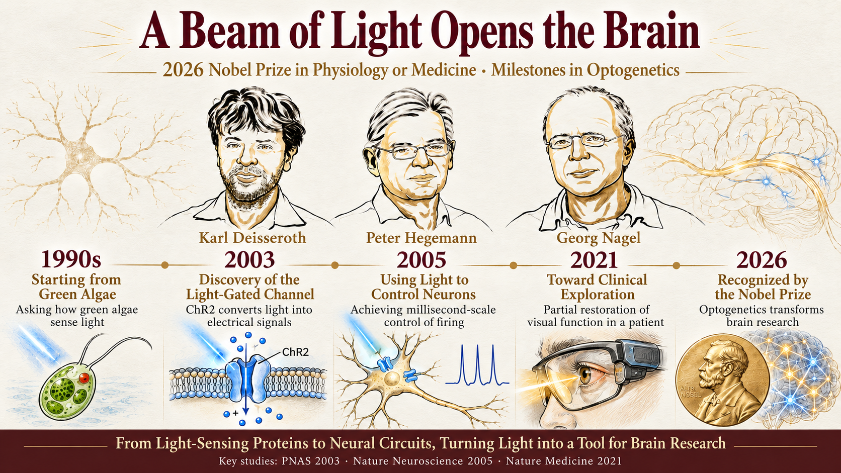
Karl Deisseroth, Peter Hegemann, and Georg Nagel received the prize for discoveries involving light-gated ion channels and optogenetics. The Nobel Committee highlighted a technology that has fundamentally changed how neuroscience is studied: using light to control the activity of specific cells.
It may sound abstract.
But more than two decades of research can be summarized in one idea:
Scientists first equipped cells with a light-responsive “switch,” then began learning how to activate that switch with increasing precision.
That second challenge remains an important direction for optogenetics today.
It Began with a Green Alga That Followed Light
The story of optogenetics did not begin in the brain. It began with a single-celled green alga capable of swimming toward a light source.
Researchers including Peter Hegemann and Georg Nagel identified a distinctive light-sensitive protein in the alga: channelrhodopsin.
Its defining feature is that light can directly control the ion channel formed by the protein.
When illuminated at an appropriate wavelength, the channel opens and charged ions cross the cell membrane, altering its electrical activity.
In 2003, Nagel and colleagues further demonstrated that channelrhodopsin-2 (ChR2) is a directly light-gated cation channel.
A pivotal step for neuroscience came in the following years.
In 2005, Deisseroth and colleagues applied ChR2 to mammalian neurons, demonstrating that optical stimulation could control neuronal firing with millisecond-scale temporal precision.
A light-sensitive protein originally found in green algae had become a tool for studying the brain.
Previously, when a group of neurons became active during a behavior, researchers could generally conclude only that the activity might be associated with that behavior.
With optogenetics, scientists could actively turn specific neurons on or off and then test whether behavior changed.
The research question shifted from:“What happened?”
to:“What happens if I change it?”
This shift from observing correlations to actively manipulating systems and testing causality is a central contribution of optogenetics. In its discussion of the 2026 Nobel Prize, Karolinska Institutet also emphasized the importance of this approach for probing causal relationships among neural circuits, memory, emotion, and behavior.
Once Cells Have a Light Switch, Where Should the Light Go?
In principle, optogenetics might seem straightforward:Express a light-sensitive protein in target cells, shine light on them, and observe the result.
In practice, laboratory experiments are considerably more complex.
The brain and other biological systems are not made up of just one cell.
A microscope field of view may contain dozens, hundreds, or more cells. Even if all of them are capable of responding to light, the experiment may focus on only a few.
The question therefore evolves from:“Can light control cells?”
to:“Which cells should be illuminated?”
As a 2013 review in Nature Neuroscience put it, realizing the full potential of optogenetics requires delivering light to the right neurons at the right time.
That need helped bring Structured Light Projection technologies, including Digital Micromirror Devices (DMDs), into optogenetics research.
From Switching on a Lamp to Programming Light
Conventional illumination is rather like turning on a lamp.
Once switched on, a broad region is illuminated.
Many modern optogenetics experiments instead require something more selective:
Light should reach only the positions specified by the researcher.
A Digital Micromirror Device (DMD) provides one way to achieve this.
Think of a DMD as an array of tiny mirrors that act like individually controllable optical switches. By adjusting their states, researchers can define the spatial pattern of projected light.
A single beam can thus become a circle, a line, or multiple distinct regions—and the pattern can change dynamically over time.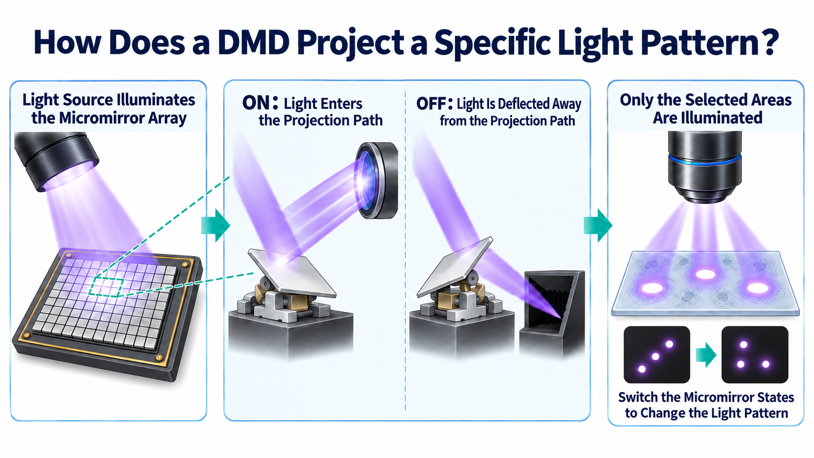
A study published in Nature Protocols in 2012 demonstrated the use of a DMD to generate arbitrary spatiotemporal light patterns for optogenetic stimulation of neurons at different positions.
By 2025, researchers had developed DMD-based stimulation systems with micrometer-scale spatial resolution and submillisecond temporal resolution, using dynamically changing illumination patterns to manipulate neuronal populations in the mouse cortex.
In effect, experimental instruments are transforming a simple act of illumination into:
Programmable light.
OptoNeuroBot®: Bringing Programmable Light into Experiments
If channelrhodopsin gives a cell a light-responsive “switch,” OptoNeuroBot® addresses the next question: how to actuate that switch with greater spatial and temporal flexibility.
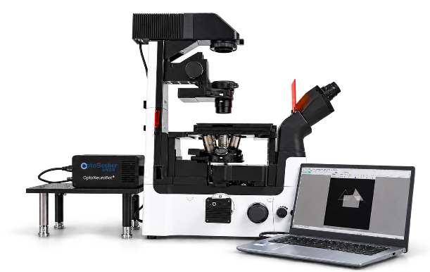
OptoNeuroBot® Structured Light Projection system integrated with a microscope
Based on DMD projection technology, OptoNeuroBot® delivers programmable spatiotemporal illumination under a microscope and supports structured light projection at subcellular scales.
Researchers can define regions of interest (ROIs) directly on microscope images, project light onto the selected regions, and control pattern shape, start time, exposure duration, and intensity. The system also supports simultaneous illumination of multiple ROIs.
Importantly, it is not designed to operate in isolation.
OptoNeuroBot® can be integrated with common upright or inverted microscopes and used alongside experimental setups such as patch-clamp recording systems and two-photon microscopy.
The prize-winning discoveries made cells responsive to optical control; Structured Light Projection systems help researchers determine more precisely where and when that light is delivered.
What Can More Precise Light Control Enable in Optogenetics?
The practical value of precise optical control is the ability to design more selective intervention experiments.
In optogenetics, researchers often want to go beyond asking whether a broad brain region matters. They also want to know:
Which cells are responsible? How do cells coordinate with one another? How do different sequences of activation change the outcome?
Using DMD-based Structured Light Projection, researchers can select one or more ROIs within the microscope field of view using OptoNeuroBot®, adjust illumination position, pattern, timing, and intensity, and combine stimulation with microscopy or patch-clamp measurements in a connected workflow:
Observe targets → Deliver targeted stimulation → Record responses
For neural circuit research, this enables targeted manipulation of selected cells or localized regions, followed by observation of other neurons’ responses, helping investigate functional connectivity and causal relationships among cells.
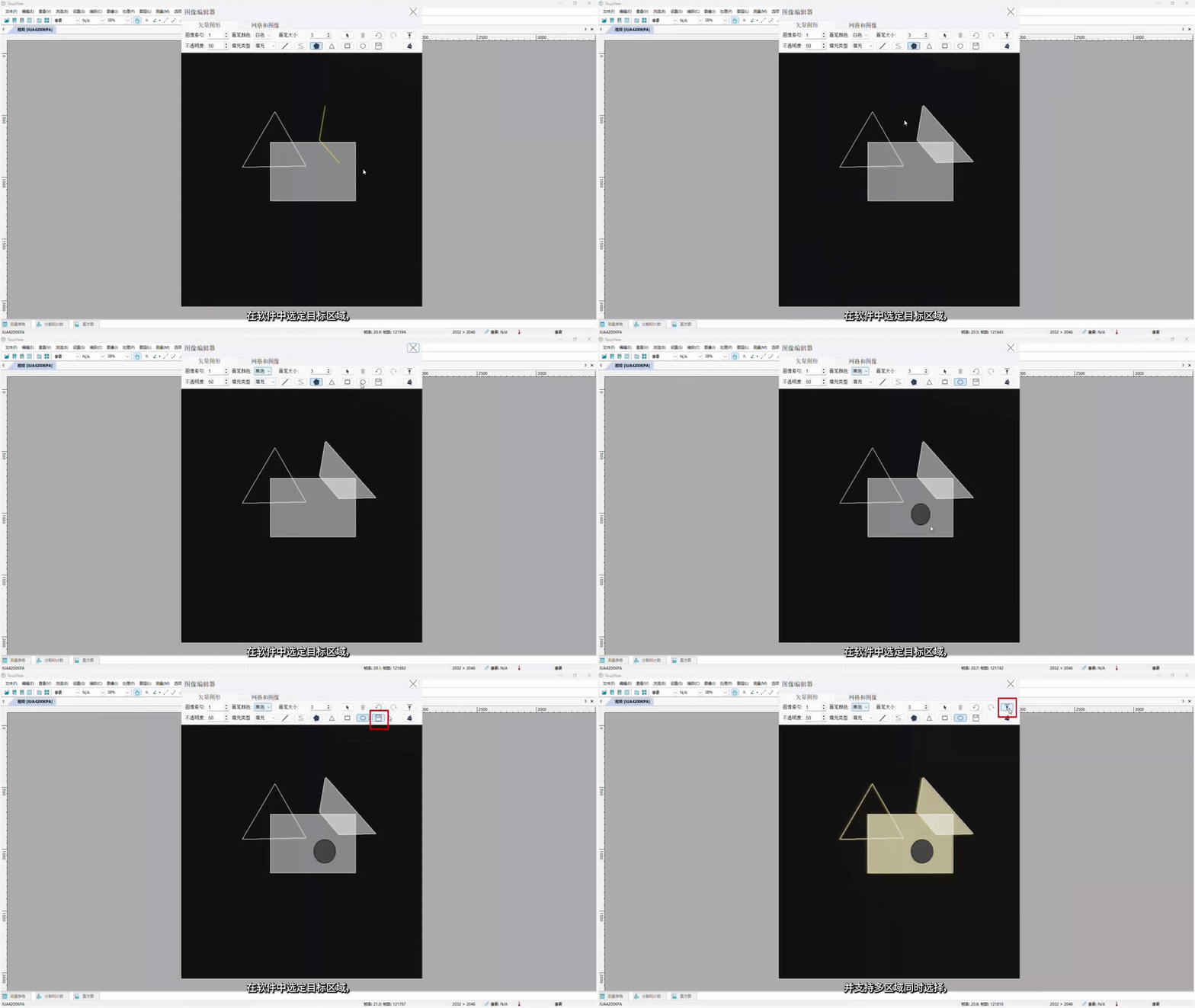
Drawing illumination spots and editing patterns in OptoNeuroBot®
Researchers can also preconfigure ROIs, stimulation sequences, and illumination patterns, then repeat experiments under matched settings—making light stimulation a programmable, controllable, and reproducible experimental variable.
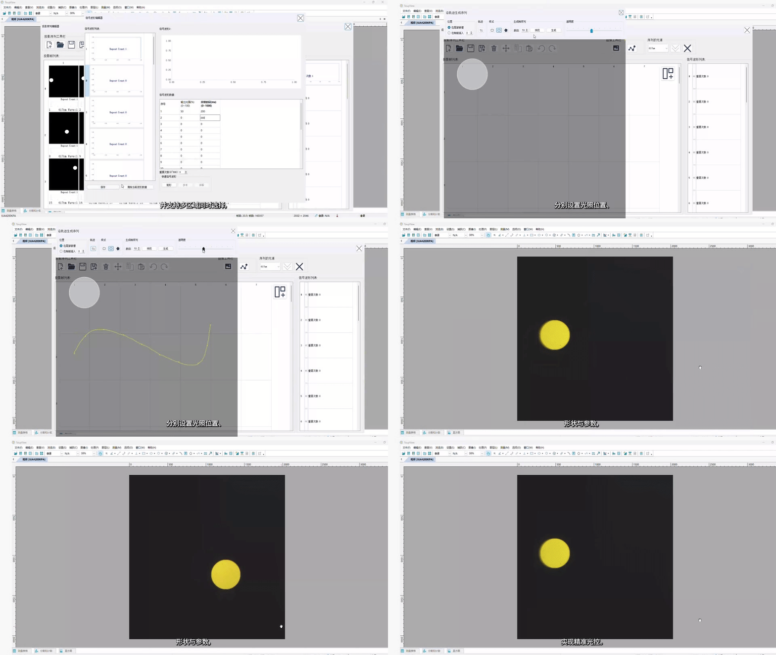
Multi-ROI combines separate target regions into one projected pattern, illuminating them simultaneously within the same projection frame.
The value of OptoNeuroBot® therefore extends beyond simply projecting light more accurately.
It helps move optogenetics from using light to activate cells toward designing interventions with light, turning optical manipulation into an executable and testable experimental protocol.
What Else Can Programmable Light Do?
Optogenetics illustrates a broader principle: when illumination position, pattern, timing, and intensity can be controlled, light itself becomes a programmable experimental tool.
This capability is not restricted to neurons. Wherever an experiment requires illumination at a designated location and time, Structured Light Projection can make experimental conditions more precisely defined and reproducible.
In micropatterning and photocuring, researchers often need to create holes, lines, gradients, or complex topological patterns in photosensitive materials for biomaterials, biomimetic substrates, cellular microenvironments, or organoid culture. OptoNeuroBot® can project user-defined patterns, combine multiple regions via Multi-ROI, and control illumination intensity and duration. Researchers can revise digital patterns and rerun experiments without repeatedly fabricating physical photomasks, potentially reducing time and cost.
In longer-term live-cell experiments, precise illumination is only one part of the workflow. After cells are removed from an incubator, changes in temperature, CO₂, and humidity can affect cell state and confound interpretation of responses to optical stimulation.
In such settings, OptoNeuroBot® can work alongside the OptoGrow® on-stage live-cell incubation system and microscope imaging. OptoNeuroBot® controls where and when light is delivered; OptoGrow® maintains suitable culture conditions; and the microscope records cellular responses. Together, they support extended single-region or multi-region stimulation and dynamic observation while helping reduce variability associated with environmental fluctuations.
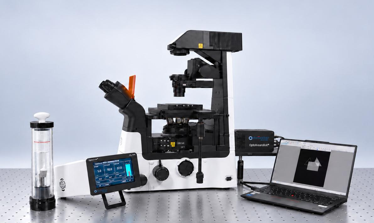
OptoNeuroBot® used with the OptoGrow® on-stage live-cell incubation system
Looking beyond optogenetics, the core capability of OptoNeuroBot® can be expressed simply:
It turns illumination from a background condition into an experimental variable that can be designed, positioned, timed, and repeated.
From Illuminating Cells to Asking Biological Questions with Light
More than two decades ago, researchers studying a green alga that swims toward light discovered ion channels that could be directly controlled by illumination.
Soon afterward, light could be used to activate specific neurons with remarkable temporal precision.
Today, with optogenetics established as a major tool in life sciences, new questions continue to emerge:
Can light be directed to selected cells alone?
Can illumination follow a spatial pattern we define?
Can different regions be stimulated in a precisely controlled sequence?
When illumination position, pattern, timing, and intensity are controllable, light does more than help us observe life. It becomes an experimental language for actively intervening in biological systems and investigating causality.
From light-gated ion channels to optogenetics and increasingly precise spatiotemporal light control, scientists have been learning to ask biological questions with that language.
OptoNeuroBot® addresses one concrete challenge within that broader effort:
Helping each experimental beam of light reach the location the researcher intends.
As scientists continue to understand life through light, OptoSeeker continues its pursuit of light.
References
1.Karolinska Institutet. Overview of the 2026 Nobel Prize in Physiology or Medicine and the award-winning discoveries.
2.Boyden ES, Zhang F, Bamberg E, Nagel G, Deisseroth K. Millisecond-timescale, genetically targeted optical control of neural activity. Nature Neuroscience, 2005.
3.Zhu P, et al. High-resolution optical control of spatiotemporal neuronal activity patterns in zebrafish using a digital micromirror device. Nature Protocols, 2012.
4.Packer AM, Roska B, Häusser M. Targeting neurons and photons for optogenetics. Nature Neuroscience, 2013.
5.Optogenetics for light control of biological systems. Nature Reviews Methods Primers, 2022.