In fields such as environmental monitoring, disease diagnosis, and drug development, the rapid and accurate analysis of biological samples is crucial. However, traditional analysis methods often face the following challenges: samples are susceptible to contamination, and manual operations are prone to errors; high reagent consumption is a disadvantage, especially for the analysis of precious biological samples; the operational process is cumbersome, and sample preparation and detection often require separate steps, leading to low efficiency.
I.Innovative Platform: The Integration of Digital Microfluidics and Raman Spectroscopy
Recently, a team from Beijing Institute of Technology published a breakthrough solution in "Biosensors and Bioelectronics": the world's first integrated digital microfluidic (DMF) and Raman spectroscopy chip.
Integrated Operation and Micro-consumption: This platform can complete the entire process from sample pre-treatment to detection on a single chip device. A significant advantage is that the entire analysis process can be completed with only 5 microliters of sample.
The Core Function of Digital Microfluidic Technology (DMF): By applying an electric field to an electrode array, it is possible to achieve precise control of micro-scale liquid droplets (such as mixing, splitting, and moving). This method significantly reduces reagent consumption and increases the level of automation.
Advantages of Raman Spectroscopy and Integration Challenges: Raman spectroscopy can provide "fingerprint" information of substance molecules, enabling non-destructive and highly specific detection. However, its inherently weak signal intensity and the difficulty of achieving effective integration into miniature devices are challenges in this field.
Core Breakthrough: Integration of the TRES (Translucent and Stacked Enhanced) Sensor: The key innovation of this research is the seamless fusion of DMF and Raman spectroscopy, with the introduction of the TRES sensor, thereby creating a highly integrated in-situ bioanalysis platform (Figure 1A-C).

Figure 1. Integrated system combining Raman spectroscopy with DMF for biochemical analysis1.
II.Research Methods: Smart System Design and Core Innovations
1. System Architecture: Precision Integrated Design
The entire platform consists of two core components:
· Digital Microfluidic (DMF) Chip: Composed of a precisely processed electrode array used to control droplet movement (Figure 1A). The chip cavity height is designed to be 100μm to ensure the precision and stability of droplet manipulation.
· TRES Sensor Integration: The TRES sensor is directly integrated onto the upper plate of the DMF chip (Figure 1B). Its specific hydrophilic structure can efficiently adsorb and enrich the target analytes onto the detection area surface, thereby improving signal acquisition efficiency.
· TRES Sensor Manufacturing Process (Figure 1D): The preparation process is simple and efficient. The glass substrate's roughness is controlled by mechanical processing, followed by the deposition of chromium and gold metal films, and finally, a UV light curing process is used to define hydrophilic and hydrophobic functional areas. It is low-cost and easy to mass-produce, making it suitable for large-scale production and disposable applications.
2. Sensor Optimization: The Exquisite Balance of Science and Engineering
The detection performance of the TRES sensor (Surface-Enhanced Raman Scattering, SERS effect) is mainly controlled by two key parameters: its surface roughness (Ra value) and the thickness of the gold film.
· Optimization of Roughness (Ra) (Figure 2): Characterization by scanning electron microscopy (SEM, Figure 2A) and white light interferometry surface profilometry (Figure 2B-C) confirmed that when the Ra value is controlled at 0.9μm, the surface micro/nanostructures are the most dense, and the hydrophilicity is the strongest, which is most favorable for analyte enrichment.
· Optimization of Gold Film Thickness (Figure 2E): Experimental data showed that when the gold film thickness is 55nm, it can most effectively excite the Surface Plasmon Resonance (SPR) effect, increasing the Raman signal intensity of the probe molecule Rhodamine B at the characteristic peak of 1505 cm⁻¹ by 1.67 times (compared to the unoptimized gold film).
· Optimization Effect (Figure 2F-G): The combination of an Ra value of 0.9μm and a gold film thickness of 55nm achieved a significant 26.3% improvement in detection sensitivity compared to the unoptimized structure. The mechanism of action (Figure 2H) is that the incident laser propagates through the glass substrate (with a refractive index higher than air), which can more efficiently couple energy to the SPR field excited by the rough surface of the gold film, thereby greatly enhancing the Raman scattering intensity of the molecules adsorbed in that area (i.e., the SERS effect).

Figure 2. Optimization of the TRES sensor1. (A) SEM images of sensors with different manufacturing parameters2. (B) White light interferometry surface profilometry and (C) roughness evaluation of the TRES sensor3. (D) Raman intensity of Rhodamine B measured from TRES sensors with different roughness and (E) gold film thicknesses4. Side view: The Raman spectrum peak is located at 1505 cm⁻¹5. (F) Comparison of dry spectra and TRES structure spectra at an Ra value of 0.9 μm6. (G) Overall enhancement effect of TRES structures with different manufacturing parameters (Intensity@1505cm⁻¹, p=0.0006)7. (H) Schematic diagram of the TRES enhancement effect8.
A Brief Analysis of the Scientific Principle: When the laser passes through the glass substrate (Figure 2H), its refractive index is higher than that of air, allowing it to couple light energy more efficiently and further amplify the SERS signal. This makes the TRES sensor stack 26.3% more sensitive than ordinary structures.
3.Construction of an Automated Experimental Platform
The entire detection system is integrated with a 3D-printed detection box, a PCB control circuit board, and an electric mobile platform (Figure 3B). After the sample (e.g., Rhodamine B solution) is diluted on the chip, the DMF system precisely controls the droplet to move to the TRES sensor detection area, and the integrated or coupled Raman spectrometer immediately performs in-situ data acquisition. The entire operation process is free from human intervention, which minimizes the risk of sample contamination.
III.Experimental Results: High-Performance Validation and Practical Applications
1. On-Chip Evaluation: A Victory for Sensitivity and Stability
Using Rhodamine B as the analyte, the system demonstrated excellent performance:
· High Sensitivity and Linearity: Within the range of 10⁻⁷ - 10⁻³ mol/L (Figure 3C), the detection signal showed a strong linear relationship with concentration (R² = 0.985). The detection limit reached 1.79 × 10⁻⁸ mol/L, comparable to professional laboratory equipment.
· Excellent Stability: The signal deviation of the 70 detection points on the sensor surface was only 7.6% (Figure 3E), which proves its spatial uniformity and ensures reliable results.
Figure 3. On-chip sample handling and detection. (A) Schematic diagram of DMF chip sample handling and TRES sensor signal detection. (B) Images of the loaded analytes on the DMF chip (top and bottom views). (C) On-chip sensitivity and linearity tests of Rhodamine B at different concentrations. (D) The sensor's calibration curve. (E) The peak values of the SERS signal measured at different parts of the TRES sensor (@1505 cm⁻¹), indicating its good spatial uniformity.
2. Biomedical Applications: In-Situ Enrichment and Detection of Exosomes
· Magnetic Bead Enrichment and Capture: Functionalized and modified magnetic beads specifically bind to and capture exosomes.
· Target Release: Exosomes are released from the surface of the magnetic beads using chemical reagents such as ammonia.
· TRES Detection: The released exosomes are efficiently adsorbed by the hydrophilic surface of the TRES sensor for Raman spectroscopy detection.
· Detection Results (Figure 4D): The obtained Raman spectrum clearly showed the characteristic peaks of exosomes, such as 963 cm⁻¹ (corresponding to lipid molecule vibrations) and 1506 cm⁻¹ (corresponding to DNA/RNA-related structures), successfully achieving specific identification of exosomes.
· Verification of Serum Sample Applicability: Although the complex composition of real serum samples causes background interference, leading to a decrease in the signal-to-noise ratio compared to exosomes suspended in a buffer solution, the system can still achieve a practical signal-to-noise ratio of 0.71, proving its potential for use in complex clinical matrices. The platform also showed a good linear response in concentration gradient experiments (Figure 4E). In addition, the system has been successfully applied to the detection of small molecules such as vitamins and antibiotics, validating its wide applicability (Figure 4E Note).

Figure 4. On-chip enrichment and detection of exosome samples1. (A) The main detection sites of exosomes2. (B) Schematic diagram of on-chip enrichment and exosome detection3. (C) Size distribution of exosomes from SEM images and their morphology on the sensor surface4. (D) Raman spectra of exosomes enriched from supernatant and serum samples using a DMF device and detected with an integrated TRES sensor5. The residual supernatant was similarly tested to evaluate the effect of non-exosomal components, and a baseline was established using a substrate without any sample as a reference6. (E) Linear fitting curve of exosome samples at different concentrations using the Raman peak at 963 cm⁻¹7
IV. Platform Advantages and Research Significance
· Highly Integrated Design: This platform successfully integrates the sample preparation steps and Raman spectroscopy detection, which are traditionally performed separately, into a single small chip device (Figures 1, 3), shortening the process by more than 80%.
· Cost-Effectiveness and Scalability: Based on its simple and efficient manufacturing process (Figures 1D, 2D), the TRES sensor has the potential for large-scale production, and the cost per unit is controllable, which is beneficial for promoting the widespread use of this technology.
· Broad Application Prospects:
o Point-of-Care Testing (POCT): Especially suitable for rapid screening of infectious diseases in resource-limited or field environments.
o Environmental Monitoring: To achieve in-situ, real-time, and high-sensitivity analysis of pollutants in environmental matrices such as water bodies.
o Drug Development: For activity testing and interaction research of trace drug molecules.
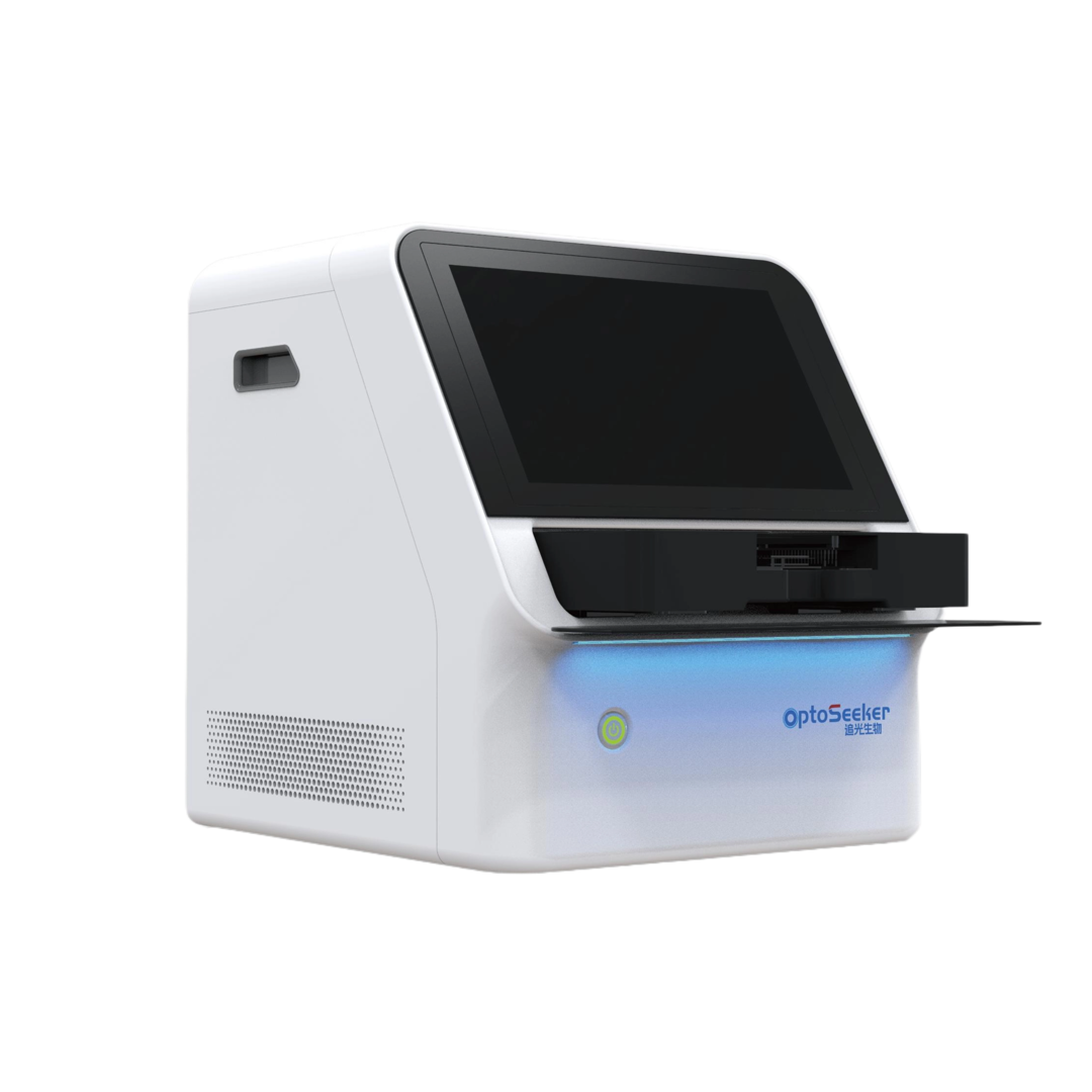
DropletBot®Digital Microfluidic Platform
V.Conclusion: Towards a Future of Smart Bioanalysis
This research not only verifies the technical feasibility of integrating digital microfluidics (DMF) and Raman spectroscopy onto a single chip platform for high-sensitivity in-situ bioanalysis but also achieves "high sensitivity, low consumption, and automation" in-situ detection through the TRES sensor. Whether for exosomes or small molecule pollutants, the system demonstrates powerful performance. With technological optimization, this platform is expected to become a standard tool for home healthcare and field research, making accurate detection readily accessible.
Article information:Wenbo Dong,Rongxin Fu,Nan Zhang,Jing Zhao,Yudan Ma, Han Cui,Jiangjiang Zhang,Zipeng Zhao,Hang Li,Yunxia Zhao,Yao Lu,Zhizhong Chen,Tianming Xu,Huikai Xie,Qian Yu,Shuailong Zhang,“Digital microfluidics with integrated Raman sensor for high-sensitivity in-situ bioanalysis”,Biosensors and Bioelectronics, 271 (2025) 117036
View Full Article:https://doi.org/10.1016/j.bios.2024.117036


