Home
Products
Optoelectronic Tweezer System
OptoBot®800 Series High-Throughput Single-Cell Optofluidic Sorting System
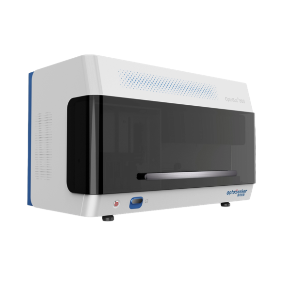
OptoBot®800 Series High-Throughput Single-Cell Optofluidic Sorting System
Optoelectronic Tweezer System
The system integrates optoelectronic tweezer (OET) technology with a fully automated microfluidic chip platform to deliver high-throughput cell screening, reducing labor and consumable costs while shortening experimental timelines from 2–3 months to 1–2 weeks.The system supports on-chip in situ culture together with two-well-plate incubation, enabling fully automated workflows from end to end. An intuitive graphical interface with configurable process steps makes custom assay design straightforward.
Product Features
- Compatible with multiple OptoTrap Chip® formats
- Automated sample loading/unloading with integrated sample needle washing
- Fully automated on-board incubation, imaging, analysis, and optoelectronic tweezer (OET) operation
- Full-chip optical imaging coverage with real-time image acquisition & storage
- Automated optical calibration
- Integrated operation and analysis software suite, including image processing and assay readout analytics
- Onboard cell culture system accommodating one 96-well plate, with 4–40 °C temperature control and a 5% CO₂ atmosphere
- Reagent bottle presence detection, automated reagent & sample level monitoring, and consumable replacement alerts
- Automated needle clogging detection with system fault alerts
- LED light source operating hours monitoring with replacement alerts


-1.png")
Areas of Interest
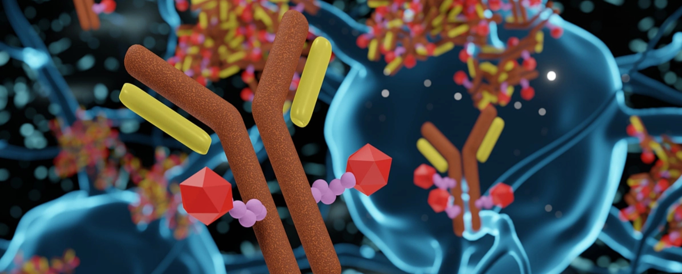

Antibody Discovery
The process isolates single B cells and validates the function of their secreted antibodies to screen for high-specificity candidates. Subsequent sequencing and bioinformatics analysis align sequencing data with functional data, enabling the identification of specific antibody sequences.
Cell and Gene Therapy (CGT)
A high-throughput, automated workflow isolates target single cells from transient co-cultures for downstream analysis, enabling multidimensional characterization of T cell potency at single-cell resolution. Images captured during isolation allow for the tracking of specific cells or cell types.
Product Comparison
Configuration Comparison Table
|
Model |
OptoBot® 1000 |
OptoBot® 800 |
|
Appearance |
 |
|
|
Product Positioning |
Fully Automated Full-size Integrated System | Benchtop System |
|
Primary Applications |
Antibody Discovery、T-cell Characterization、Cell Line Development | Antibody Discovery、T-cell Characterization |
|
Hardware Design |
Floor-standing / All-in-one Integrated System; Includes Integrated Touchscreen, PC, and Keyboard |
Benchtop design; connects to an external workstation |
|
Pneumatic Configuration |
GMP standards; Supports Positive Pressure (Express) or Negative Pressure (Standard/Plus) configurations | GMP standards |
|
Automated Incubation |
On-chip in situ incubation + two 96-well plates | On-chip in situ incubation + one 96-well plate |
|
Incubation Control |
Temperature: 10–50 °C (on-chip) / 4–40 °C (plate); 5% CO₂ | Aligned with OptoBot® 1000 |
|
Optics & Imaging |
5 Fluorescence Channels + Brightfield; |
Aligned with OptoBot® 1000 |
|
Objective Lenses |
Standard: 4X, 10X; Optional: 20X | Aligned with OptoBot® 1000 |
|
Sample I/O |
1.5 mL Eppendorf (EP) tubes, 0.2 mL PCR tubes, standard 96-well microplates / PCR plates | Aligned with OptoBot® 1000 |
|
Target Cell Types |
CHO cells, plasma B cells, memory B cells, T cells, hybridomas, primary cells, adherent cells, etc. | Aligned with OptoBot® 1000 |